Download
1 / 44
450 likes | 539 Vues
Unveil the mysteries of E. coli bacteria, from its beneficial roles in digestion to the dangerous strains causing illnesses. Learn about the good and bad aspects of E. coli, its sources of contamination, and how it affects the human body. Discover the science behind E. coli toxins and the mechanisms of infection. Delve into the world of bacteria with informative visuals and intriguing facts.

E N D
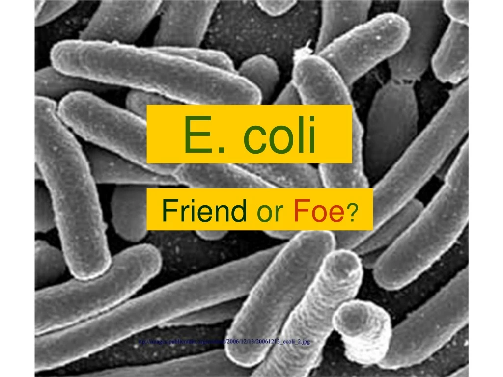
E. coli FriendorFoe?
Why, there are stories all over… Let’s see what that book is..
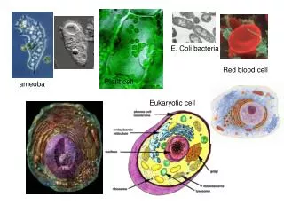
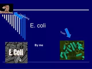
E. coli (Escherichia coli) • Gram negative, rod-shaped bacteria • Lives in the intestine of vertebrates • Anaerobic (lives without oxygen) • Despite one species, there are many different strains (many different DNA sequences) • All have a similar life cycle transmission So what is E. coli? Looks like a bug to me…
E. coli: The Good E. coli Benefits • Symbiotically coexist with us • E. coli gets food and shelter • We get the vitamins (K and B complexes) E. coli produce and help in breaking down food • E. coli help digest certain food materials. E. coli within the villi TGI E. coli (Thank God Its E. coli)
Hundreds of strains of E. coli exist I love chocolate peanut butter It is kind of like ice cream Some Good. Some Bad. Asparagus flavored ice cream… Yuk!
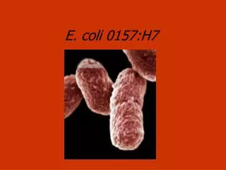
Let’s focus on the bad guys E. coli: The Bad Some Bad Strains of E. coli • E. coli O157:H7 • E. coli O104:H21 • E. coli O121 • E. coli O111:NM • E. coli O113:H21 The O’s and H’s, and NM’s and numbers refer to the antigens found on the bacteria. Anything you say…
One bad strain is E. coli 0157:H7 E. coli: The Bad • Emerging disease-causing strains of E. coli identified in early 1980’s • Can cause bloody diarrhea and severe stomach cramps • Some strains of E. coli 0157:H7 make toxins (called shiga-like) symbolized Stx2 • Can cause serious kidney impairment (sometimes resulting in death) Oh.. no.. I want to say… I hope they don’t have E-coli O157:H7
O:157:H7 E. coli: The Bad I became that way because of … Click on me if you dare… Do we have a choice?
One Hypothesis E. coli: The Bad • The bad gene--E. coli O157:H7 Toxin 2 gene--is on a bacteriophage • Bacteriophages (phages) are viruses that infect bacteria. • Phages can insert their DNA into the bacterial DNA Diagram of a bacteriophage Looks like some bad mosquito Compliments of http://oceanworld.tamu.edu/resources/oceanography-book/Images/BacteriophageCartoon.jpg
Phages infecting E. coli. DNA is injected into the cell. The outer protein structure stays behind. E. coli: The Bad . E. coli O157:H7-- a good bacteria gone bad… So how do we get this bad bacteria? Compliments of : http://www.vet.upenn.edu/schoolresources/communications/publications/bellwether/55/images/bacteriophage.jpg
Sources of E-coli Contamination Sources of E. coli Contamination http://www.umassvegetable.org/images/soilsr_cops_pest_mgt/crop/lettuce3.jpg http://www.hornimports.com/images/perth-show-oliver1.jpg E. coli is not just on the outside of the plants, but also internal, contained vascularly as well http://meat.tamu.edu/images/metmyoglobin2.jpg http://images.usatoday.com/news/_photos/2006/09/14/spinach.jpg So...Ground beef, plants such as spinach and lettuce, even raw milk could potentially be tainted with E. coli O:157:H7… The newspaper articles are beginning to make more sense.
Drinking water or swimming in water contaminated with fecal material Hmm…So that is why they made us get out of the pool when little Billy had the problem… Sources of E. coli Contamination
Wait… Is this sign I saw in the bathroom related? Preparing food without properly washing your hands…. Sources of E. coli Contamination
http://www.fair-safety.com/related-sites.htm E. coli in the Body Some E. coli, such as E. coli O157:H7, release powerful toxins. Click on the image on the right to see a movie which indicates what happens in the intestinal tract… (Be certain your sound is on…) Movie clip compliments of http://www.fair-safety.com/ecoli.mov Ooh…I don’t know if I am prepared for this
E. coli O157:H7 and other bad E. coli strains have a toxin that damages the intestinal walls. But it goes beyond that too… Here is a model showing the atoms making up the Stx2 toxin–that’s it name-- released by E. coli O157:H7 Carbon atoms are the grey spheres, oxygen the red spheres, nitrogen the bluespheres, and sulfur the yellow spheres E. coli in the Body
These two models are representing the same toxin produced by E. coli O157:H7 Space-filled model So those two are the same… that’s wild Backbone model E. coli in the Body
The same E. coli O157:H7 toxin, only rotated. Blue part = cell membrane binding piece Red part = piece that binds to a part of the ribosome It’s kind of…pretty. I like the star shape E. coli in the Body
Key Port of Entry: Glycosphingolipid Globotriasosylceramide Gb3 for short These molecules are naturally found embedded within the cell membranes of various animals in varying quantities. Some cells (kidney) have lots of these molecules. Others cells or animals do not. (Cows and pigs do not have Gb3) Say that 3 times… E. coli in the Body
People have found that the E.coli toxin attaches to Gb3 located in the cell membrane (The red part of the toxin that binds to the ribosome is not shown here.) Whoever first saw this, must have said this was pretty cool! E. coli in the Body
The E. coli O157:H7’s toxin, called Stx2, binds moreeffectively than other strains’ toxins to the Gb3 because of differences it has at its various binding sites. To see a comparison the three E. coli O157:H7 Stx2 toxin’s binding domains and a different pathogenic E. coli toxin, click here. …Ooh…. E. coli in the Body
Receptor Binding Site 1 Note the green residue groups pictured in the grooves are different. These groups attach to the Gb3 receptor. Toxin from less virulent strain of E.coli called Stx1 Stx2 toxin I think they mean here E. coli in the Body
Receptor Binding Site 2 Differences in the size of the attractive groups can be seen here. Stx1 Toxin Stx2 toxin It’s amazing people can detect differences at the atomic level, isn’t it? E. coli in the Body
Receptor Binding Site 3 This binding site is fairly similar between strains… Stx1 Toxin Stx2 Toxin Looks the same to me E. coli in the Body
Close-up of binding differences between E. coli O157:H7 and the Stx toxin by some other E. coli. Look at how the yellow and green groups lie with respect to each other… Receptor Binding Site 3 That would mean that one of the toxins would bond more strongly, right? Which one? E. coli in the Body
The toxin is attracted to the Gb3 sites in the membrane and sticks to the Gb3 receptors Cell membrane of blood vessel or intestinal tract In the intestinal tract You didn’t really want to see the stuff the hamburger is processed into… Or show the end product E. coli in the Body
The toxin is then taken into the cell by receptor-mediated endocytosis There is a lot of stuff in there, Isn’t there? E. coli in the Body
Golgi Body The toxin presumably makes its way from the endosome to a Golgi body to the endoplasmic reticulum Very Rough Endoplasmic Reticulum What a ride! E. coli in the Body
Somewhere in the endoplasmic reticulum, it is believed the toxin breaks into two different part; the pieces being released from each other are destined to go bouncing around in the cytoplasm Active-Binding Site It doesn’t look like people really know the exact process, yet… Complete Toxin Part of A and receptor binding site E. coli in the Body
On the 28 S ribosome is an adenine group to which the active site of the toxin is attracted. But the more real image is a bit more complicated and I don’t think we want to go there… Adenine doesn’t really sit on the ribosome like that… http://www.cartage.org.lb/en/themes/Sciences/Zoology/AnimalPhysiology/Anatomy/AnimalCellStructure/Ribosomes/ribosome.jpg E. coli in the Body
Don’t say that I didn’t warn you… Now let’s go back… Those are atoms. E. coli in the Body
Stx2 Receptor Binding Domain Eventually, the active binding piece finds itself near the 28S part of a ribosome. Oh – Oh! This doesn’t look good for Ribo (The active binding site is highlighted in yellow, and models are not to scale.) E. coli in the Body http://www.cartage.org.lb/en/themes/Sciences/Zoology/AnimalPhysiology/Anatomy/AnimalCellStructure/Ribosomes/ribosome.jpg
The adenine gets cleaved from the ribosome. Unfortunately, the ribosome needs the adenine to work properly. Stx2 Receptor Binding Domain with adenine attached I think this is going to be sad. http://www.cartage.org.lb/en/themes/Sciences/Zoology/AnimalPhysiology/Anatomy/AnimalCellStructure/Ribosomes/ribosome.jpg E. coli in the Body
As a result proteins can’t be made and the cell dies. Hence, cells with lots of Gb3 receptors (kidney, lung…) that allow the E. coli toxin in are the ones that will die. http://www.cartage.org.lb/en/themes/Sciences/Zoology/AnimalPhysiology/Anatomy/AnimalCellStructure/Ribosomes/ribosome.jpg E. coli in the Body
E. coli O157:H7 toxin, STX2, is more toxic than STX for instance, because its receptor binding domain is more open for the adenine to bond Purple is showing the part of the structure blocking the site where the adenine binds. Yellow is showing where the adenine binds. Stx2 Receptor Binding Domain Stx Receptor Binding Domain I’m okay. I’ve composed myself. Thanks for caring! E. coli in the Body
Summarizing E. coli… Let me see if I get it… E. coli O157:H7 emits a toxin that is more potent, more dangerous, more virulent (Are you proud of me using that word? ) because the toxin that E. coli O157:H7 emits, … Stx2, due to its chemical structure, has a greater ability 1. to get into the cell and, 2. once in the cell, to disable the ribosome. Is that right?
I now understand why people are concerned with E. coli O157:H7 Thanks for learning with me… If you are interested in learning more about E. coli, please check out the following web sites on the next page…
Primary Citations Research article by Structure of Shiga Toxin Type 2 (Stx2) from Escherichia coli O157:H7* Received for publication, February 22, 2004, and in revised form, April 7, 2004 Published, JBC Papers in Press, April 9, 2004, DOI 10.1074/jbc.M401939200 Marie E. Fraser‡, Masao Fujinaga§, Maia M. Cherney§, Angela R. Melton-Celsa¶, Edda M. Twiddy¶, Alison D. O’Brien¶, and Michael N. G. James§ Pdb files used for Rasmol images for Stx1 and Stx2 toxins: 1rq1 (Stx) 1rp1 (Stx2) from the Protein Data Bank http://www.rcsb.org/pdb/home/home.do Rasmol software is available from http://www.umass.edu/microbio/rasmol/
Cover image gray E.coli around the message http://images.publicradio.org/content/2006/12/13/20061213_ecoli_2.jpg Ground beef http://meat.tamu.edu/images/metmyoglobin2.jpg Lettuce http://www.umassvegetable.org/images/soils_crops_pest_mgt/crop/lettuce3.jpg Spinach http://images.usatoday.com/news/_photos/2006/09/14/spinach.jpg Milk http://www.hornimports.com/images/perth-show-oliver1.jpg Notice sign http://www.speedysigns.com/images/osha/large/NOTICE01.gif Sound effect http://www.partnersinrhyme.com/soundfx/human.shtml E.coli movie http://www.fair-safety.com/ecoli.mov Bacterophage http://oceanworld.tamu.edu/resources/oceanography-book/Images/BacteriophageCartoon.jpg http://www.vet.upenn.edu/schoolresources/communications/publications/bellwether/55/images/bacteriophage.jpg Endoplasmic Reticulum http://biodidac.bio.uottawa.ca/ftp/BIODIDAC/ZOO/CELL/DIAGBW/CELL018B.GIF Citations for images used in the presentation
Golgi Bodies http://images.google.com/imgres?imgurl=http://www.rsc-yh.ac.uk/images/screenshots/clip_image004.gif&imgrefurl=http://www.rsc-yh.ac.uk/usingword.asp&h=74&w=107&sz=3&hl=en&start=63&um=1&tbnid=mIla7J7y1vSK4M:&tbnh=59&tbnw=85&prev=/images%3Fq%3DGolgi%2BBody%26start%3D54%26ndsp%3D18%26svnum%3D10%26um%3D1%26hl%3Den%26client%3Dfirefox-a%26rls%3Dorg.mozilla:en-US:official%26sa%3DN Ribosomes http://www.cartage.org.lb/en/themes/Sciences/Zoology/AnimalPhysiology/Anatomy/AnimalCellStructure/Ribosomes/ribosome.jpg E.coli Newspaper http://www.ecoliblog.com/souplantation-infosheet.jpg http://www.ecoliblog.com/minnesota-ecoli.jpg http://seattlepi.nwsource.com/health/285378_spinach16.html http://www.cbc.ca/canada/montreal/story/2006/08/21/pools-polluted.html?ref=rss http://www.foxnews.com/story/0,2933,215157,00.html http://whyfiles.org/246e_coli/ http://news.bbc.co.uk/2/hi/uk_news/wales/4546606.stm Citations for images used in the presentation