Download
1 / 29
290 likes | 352 Vues
Explore the fascinating world of cells, from their structure to how researchers study them using microscopes and cell fractionation. Learn about the differences between prokaryotic and eukaryotic cells, the roles of the nucleus and ribosomes, and more. Join us on this educational tour of the cell! |

E N D
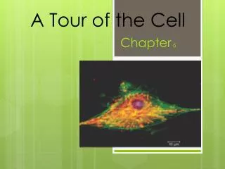
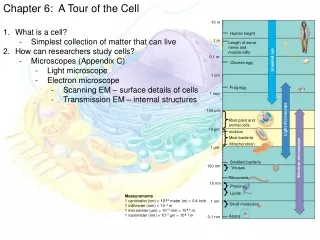
Chapter 6: A Tour of the Cell 10 m 1. What is a cell? - Simplest collection of matter that can live 2. How can researchers study cells? - Microscopes (Appendix C) - Light microscope - Electron microscope - Scanning EM – surface details of cells - Transmission EM – internal structures Human height 1 m Length of some nerve and muscle cells Unaided eye 0.1 m Chicken egg 1 cm Frog egg 1 mm Light microscope 100 µm Most plant and animal cells nucleus Most bacteria 10 µm Nucleus Most bacteria Electron microscope Mitochondrion 1 µm Smallest bacteria Viruses 100 nm Ribosomes 10 nm Proteins Lipids Measurements 1 centimeter (cm) = 102meter (m) = 0.4 inch 1 millimeter (mm) = 10–3m 1 micrometer (µm) = 10–3mm = 106m 1 nanometer (nm) = 10–3µm = 10 9 m 1 nm Small molecules Atoms 0.1 nm
Chapter 6: A Tour of the Cell 1. What is a cell? - Simplest collection of matter that can live 2. How can researchers study cells? - Microscopes (Appendix C) - Cell fractionation - Used to isolate cellular components based on size & density - Cells are homogenized - Organelles separated by differential centrifugation Homogenization Tissue cells 1000 g Homogenate (1000 times the force of gravity) 10 min Differential centrifugation Supernatant poured into next tube 20,000 g 20 min 80,000 g 60 min Pellet rich in nuclei and cellular debris 150,000 g 3 hr Pellet rich in mitochondria (and chloro- plasts if cells are from a plant) Pellet rich in “microsomes” (pieces of plasma mem- branes and cells’ internal membranes) Pellet rich in ribosomes
Chapter 6: A Tour of the Cell 1. What is a cell? 2. How can researchers study cells? 3. What is the difference between prokaryotic & eukaryotic cells? - Prokaryotic - No true nucleus or other membrane-bound organelles - Bacteria & Archaebacteria Pili: attachment structures on the surface of some prokaryotes Nucleoid: region where the cell’s DNA is located (not enclosed by a membrane) Ribosomes: organelles that synthesize proteins Plasma membrane: membrane enclosing the cytoplasm Cell wall: rigid structure outside the plasma membrane Capsule: jelly-like outer coating of many prokaryotes 0.5 µm Flagella: locomotion organelles of some bacteria (b) A thin section through the bacterium Bacillus coagulans (TEM) (a) A typical rod-shaped bacterium
Chapter 6: A Tour of the Cell 1. What is a cell? 2. How can researchers study cells? 3. What is the difference between prokaryotic & eukaryotic cells? - Prokaryotic - No true nucleus or other membrane-bound organelles - Bacteria & Archaebacteria - Eukaryotic - Nucleus & other membrane-bound organelles are present - Plants, animals, fungi, protists
Figure 6.9 Animal Cell Nuclear envelope ENDOPLASMIC RETICULUM (ER) NUCLEUS Nucleolus Rough ER Smooth ER Chromatin Flagellum Plasma membrane Centrosome CYTOSKELETON Microfilaments Intermediate filaments Ribosomes Microtubules Microvilli Golgi apparatus Peroxisome In animal cells but not plant cells: Lysosomes Centrioles Flagella (in some plant sperm) Lysosome Mitochondrion
Figure 6.9 Plant Cell Nuclear envelope Rough endoplasmic reticulum Nucleolus NUCLEUS Chromatin Smooth endoplasmic reticulum Centrosome Ribosomes ( small brown dots ) Central vacuole Tonoplast Golgi apparatus Microfilaments Intermediate filaments CYTOSKELETON Microtubules Mitochondrion Peroxisome Plasma membrane Chloroplast Cell wall Plasmodesmata In plant cells but not animal cells: Chloroplasts Central vacuole and tonoplast Cell wall Plasmodesmata Wall of adjacent cell
Chapter 6: A Tour of the Cell 1. What is a cell? 2. How can researchers study cells? 3. What is the difference between prokaryotic & eukaryotic cells? 4. What is the role of the nucleus? - Houses most of the genes on chromosomes made of chromatin - Chromatin = DNA wrapped around proteins - Surrounded by nuclear envelope - Double membrane lined with pores - Pore complexes regulate movement of RNA & proteins into & out of nucleus - Nuclear lamina – inner lining of nuclear envelope used for support - Nucleolus - Site of ribosome production - rRNA + imported ribosomal proteins (through pore complexes)
Figure 6.10 The nucleus and its envelope Nucleus Nucleus 1 µm Nucleolus Chromatin Nuclear envelope: Inner membrane Outer membrane Nuclear pore Pore complex Rough ER Surface of nuclear envelope. TEM of a specimen prepared by a special technique known as freeze-fracture. 0.25 µm Ribosome 1 µm Close-up of nuclear envelope Nuclear lamina (TEM). The netlike lamina lines the inner surface of the nuclear envelope. Pore complexes (TEM). Each pore is ringed by protein particles.
Chapter 6: A Tour of the Cell 1. What is a cell? 2. How can researchers study cells? 3. What is the difference between prokaryotic & eukaryotic cells? 4. What is the role of the nucleus? 5. What is the role of the ribosome? - Protein synthesis (aka…translation) - Made in nucleolus - Large & small subunit - Free ribosomes in cytosol - Bound ribosomes on rough ER or nucleus Ribosomes ER Cytosol Endoplasmic reticulum (ER) Free ribosomes Bound ribosomes Large subunit Small subunit 0.5 µm TEM showing ER and ribosomes Diagram of a ribosome
Chapter 6: A Tour of the Cell 1. What is a cell? 2. How can researchers study cells? 3. What is the difference between prokaryotic & eukaryotic cells? 4. What is the role of the nucleus? 5. What is the role of the ribosome? 6. What is the endomembrane system & who are its members? - Collection of membranes inside a eukaryotic cell related through direct contact or by transfer vesicles - Nuclear envelope, endoplasmic reticulum (ER), Golgi apparatus, lysosomes, vacuoles, plasma membrane 7. What is the role of the smooth ER? - Make lipids (oils, phospholipids, & steroids) - Metabolism of carbs - Detoxification of drugs & poisons - Ca+2ion storage 8. What is the role of the rough ER? - Studded with ribosomes - Makes secreted proteins, membranes & glycoproteins Smooth ER Nuclear envelope Rough ER ER lumen Cisternae Ribosomes Transport vesicle Smooth ER Transitional ER Rough ER200 µm
Chapter 6: A Tour of the Cell 1. What is a cell? 2. How can researchers study cells? 3. What is the difference between prokaryotic & eukaryotic cells? 4. What is the role of the nucleus? 5. What is the role of the ribosome? 6. What is the endomembrane system & who are its members? 7. What is the role of the smooth ER? 8. What is the role of the rough ER? 9. What is the role of the Golgi apparatus? - Center of manufacturing, warehousing, sorting & shipping - ER products get modified & sent along - Membrane phospholipids - Sugars of glycoproteins - Targets proteins for other organelles - Sorts products for secretion
Figure 6.13 The Golgi apparatus Golgi apparatus cis face (“receiving” side of Golgi apparatus) 1 2 Vesicles coalesce to form new cis Golgi cisternae Vesicles move from ER to Golgi 0.1 0 µm Vesicles also transport certain proteins back to ER 6 Cisternae Cisternal maturation: Golgi cisternae move in a cis- to-trans direction 3 4 Vesicles form and leave Golgi, carrying specific proteins to other locations or to the plasma mem- brane for secretion 5 trans face (“shipping” side of Golgi apparatus) Vesicles transport specific proteins backward to newer Golgi cisternae TEM of Golgi apparatus
Chapter 6: A Tour of the Cell 1. What is a cell? 2. How can researchers study cells? 3. What is the difference between prokaryotic & eukaryotic cells? 4. What is the role of the nucleus? 5. What is the role of the ribosome? 6. What is the endomembrane system & who are its members? 7. What is the role of the smooth ER? 8. What is the role of the rough ER? 9. What is the role of the Golgi apparatus? 10.What do lysosomes do? - Digestion & recycling at pH 5 - Hydrolytic enzymes break bonds of all macromolecules
Figure 6.14 Lysosomes 1 µm Nucleus Lysosome containing two damaged organelles 1 µ m Mitochondrion fragment Peroxisome fragment Lysosome Lysosome contains active hydrolytic enzymes Lysosome fuses with vesicle containing damaged organelle Hydrolytic enzymes digest organelle components Hydrolytic enzymes digest food particles Food vacuole fuses with lysosome Digestive enzymes Lysosome Lysosome Lysosome Plasma membrane Digestion Digestion Vesicle containing damaged mitochondrion Food vacuole (a) Phagocytosis: lysosome digesting food (b) Autophagy: lysosome breaking down damaged organelle
Chapter 6: A Tour of the Cell 1. What is a cell? 2. How can researchers study cells? 3. What is the difference between prokaryotic & eukaryotic cells? 4. What is the role of the nucleus? 5. What is the role of the ribosome? 6. What is the endomembrane system & who are its members? 7. What is the role of the smooth ER? 8. What is the role of the rough ER? 9. What is the role of the Golgi apparatus? 10.What do lysosomes do? 11. What do vacuoles do? - Food vacuoles – lysosomes - Contractile vacuoles – FW protists use these to pump out excess water - Central vacuole – reserve of many different substances or ions for plants - Let’s review the endomembrane system……
Figure 6.16 Review: relationships among organelles of the endomembrane system 1 Nuclear envelope is connected to rough ER, which is also continuous with smooth ER Nucleus Rough ER Smooth ER Nuclear envelope 3
Figure 6.16 Review: relationships among organelles of the endomembrane system 1 Nuclear envelope is connected to rough ER, which is also continuous with smooth ER Nucleus Rough ER 2 Membranes and proteins produced by the ER flow in the form of transport vesicles Smooth ER cis Golgi Nuclear envelope to the Golgi Transport vesicle 3 3 Golgi pinches off transport vesicles and other vesicles that give rise to lysosomes and vacuoles trans Golgi Lysosome available for fusion with another vesicle for digestion 4 Transport vesicle carries 5 proteins to plasma membrane for secretion
Figure 6.16 Review: relationships among organelles of the endomembrane system 1 Nuclear envelope is connected to rough ER, which is also continuous with smooth ER Nucleus Rough ER 2 Membranes and proteins produced by the ER flow in the form of transport vesicles Smooth ER cis Golgi Nuclear envelope to the Golgi Transport vesicle 3 3 Golgi pinches off transport vesicles and other vesicles that give rise to lysosomes and Plasma membrane vacuoles trans Golgi Lysosome available for fusion with another vesicle for digestion 4 Transport vesicle carries 5 proteins to plasma membrane for secretion 6 Plasma membrane expands by fusion of vesicles; proteins are secreted from cell
Chapter 6: A Tour of the Cell 1. What is a cell? 2. How can researchers study cells? 3. What is the difference between prokaryotic & eukaryotic cells? 4. What is the role of the nucleus? 5. What is the role of the ribosome? 6. What is the endomembrane system & who are its members? 7. What is the role of the smooth ER? 8. What is the role of the rough ER? 9. What is the role of the Golgi apparatus? 10.What do lysosomes do? 11. What do vacuoles do? 12.What is the role of a mitochondria? - Site of cellular respiration - Double membrane (inner & outer) & mitochondrial matrix - Has DNA & ribosomes & can reproduce on their own
Figure 6.17 The mitochondrion, site of cellular respiration Mitochondrion Intermembrane space Outer membrane Free ribosomes in the mitochondrial matrix Inner membrane Cristae Matrix Mitochondrial DNA 100 µm
Chapter 6: A Tour of the Cell 1. What is a cell? 2. How can researchers study cells? 3. What is the difference between prokaryotic & eukaryotic cells? 4. What is the role of the nucleus? 5. What is the role of the ribosome? 6. What is the endomembrane system & who are its members? 7. What is the role of the smooth ER? 8. What is the role of the rough ER? 9. What is the role of the Golgi apparatus? 10.What do lysosomes do? 11. What do vacuoles do? 12.What is the role of a mitochondria? 13.What do chloroplasts do? - Photosynthesis for plant energy transformations - Double membrane (inner & outer) & stroma - Has DNA & ribosomes & can reproduce on their own - Thylakoids have chlorophyll & harness light energy
Figure 6.18 The chloroplast, site of photosynthesis Chloroplast Ribosomes Stroma Chloroplast DNA Inner and outer membranes Granum 1 µm Thylakoid
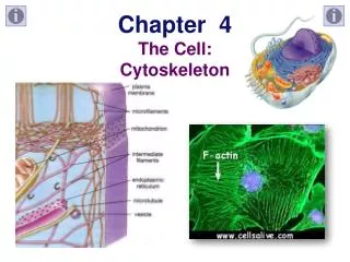
Chapter 6: A Tour of the Cell 1. What is a cell? 2. How can researchers study cells? 3. What is the difference between prokaryotic & eukaryotic cells? 4. What is the role of the nucleus? 5. What is the role of the ribosome? 6. What is the endomembrane system & who are its members? 7. What is the role of the smooth ER? 8. What is the role of the rough ER? 9. What is the role of the Golgi apparatus? 10.What do lysosomes do? 11. What do vacuoles do? 12.What is the role of a mitochondria? 13.What do chloroplasts do? 14.What about peroxisomes? - Enzymes that transfer hydrogen to oxygen forming peroxide - Beta oxidation of fatty acids for energy - Detoxifying alcohol in liver 15.How does the cell keep its shape? - Cytoskeleton made of - Microtubules - Microfilaments - Intermediate filaments - Role – support, motility & regulation
Chapter 6: A Tour of the Cell 1. What is a cell? 2. How can researchers study cells? 3. What is the difference between prokaryotic & eukaryotic cells? 4. What is the role of the nucleus? 5. What is the role of the ribosome? 6. What is the endomembrane system & who are its members? 7. What is the role of the smooth ER? 8. What is the role of the rough ER? 9. What is the role of the Golgi apparatus? 10.What do lysosomes do? 11. What do vacuoles do? 12.What is the role of a mitochondria? 13.What do chloroplasts do? 14.What about peroxisomes? 15.How does the cell keep its shape? 16.What important structures lie outside of the cell? - Plants – primary cell wall (initially thin & flexible) & secondary cell wall in mature cells - Animals – Extra cellular matrix (ECM) – glycoproteins from cell - Collagen embedded in proteoglycans - Fibronectin & integrins
Figure 6.29 Extracellular matrix (ECM) of an animal cell Collagen fibers are embedded in a web of proteoglycan complexes. A proteoglycan complex consists of hundreds of proteoglycan molecules attached noncovalently to a single long polysac- charide molecule. Polysaccharide molecule EXTRACELLULAR FLUID Carbo- hydrates Fibronectin attaches the ECM to integrins embedded in the plasma membrane. Core protein Integrins are membrane proteins that are bound to the ECM on one side and to associated proteins attached to microfilaments on the other. This linkage can transmit stimuli between the cell’s external environment and its interior and can result in changes in cell behavior. Proteoglycan molecule Plasma membrane CYTOPLASM Micro- filaments Integrin
Chapter 6: A Tour of the Cell 1. What is a cell? 2. How can researchers study cells? 3. What is the difference between prokaryotic & eukaryotic cells? 4. What is the role of the nucleus? 5. What is the role of the ribosome? 6. What is the endomembrane system & who are its members? 7. What is the role of the smooth ER? 8. What is the role of the rough ER? 9. What is the role of the Golgi apparatus? 10.What do lysosomes do? 11. What do vacuoles do? 12.What is the role of a mitochondria? 13.What do chloroplasts do? 14.What about peroxisomes? 15.How does the cell keep its shape? 16.What important structures lie outside of the cell? 17.How are neighboring cells connected? - Plants – plasmodesmata – openings in cell walls that cytosol can pass through - Animals - Tight junctions – membranes of neighboring cells bound by specific proteins - Desmosomes – function like rivets fastening cells together into strong sheets - Gap junctions – cytoplasmic channels between cells
Figure 6.31 Exploring Intercellular Junctions in Animal Tissues TIGHT JUNCTIONS At tight junctions, the membranes of neighboring cells are very tightly pressed against each other, bound together by specific proteins (purple). Forming continu- ous seals around the cells, tight junctions prevent leakage of extracellular fluid across a layer of epithelial cells. Tight junction Tight junctions prevent fluid from moving across a layer of cells 0.5 µm DESMOSOMES Desmosomes (also called anchoring junctions) function like rivets, fastening cells together into strong sheets. Intermediate filaments made of sturdy keratin proteins anchor desmosomes in the cytoplasm. Tight junctions Intermediate filaments Desmosome Gap junctions 1 µm GAP JUNCTIONS Gap junctions (also called communicating junctions) provide cytoplasmic channels from one cell to an adjacent cell. Gap junctions consist of special membrane proteins that surround a pore through which ions, sugars, amino acids, and other small molecules may pass. Gap junctions are necessary for commu- nication between cells in many types of tissues, including heart muscle and animal embryos. Extracellular matrix Space between cells Gap junction Plasma membranes of adjacent cells 0.1 µm