Download
1 / 45
470 likes | 628 Vues
Explore the anatomy and physiology of single lung ventilation, understand the available techniques, and learn about the indications and contraindications for different procedures in thoracic surgery. Discover the advantages and disadvantages of various methods to choose the most suitable technique for your patient.

E N D
Dr. SOMALETHA T. BHATTACHARYA M.D. FFARCSI MASSACHUSSETS GENERAL HOSPITAL BOSTON SINGLE LUNG VENTILATION
DISCLOSURES No conflict of interest or disclosures
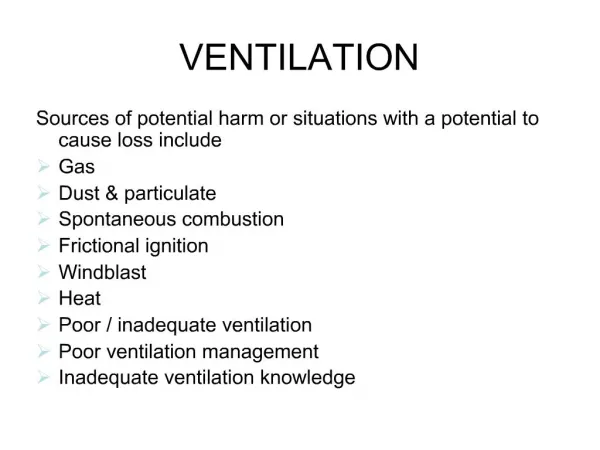
OBJECTIVES Anatomy of the airway Physiology of single lung ventilation Indications and contraindications Available techniques Advantages and disadvantages of each How to choose a particular technique for a particular patient.
ANATOMY OF THE TRACHEA Tracheal cross-section circular (infants) /ovoid (adults) A-P diameter limits the size of tube Bifurcates into right & left bronchi Right tracheobronchial angle is more acute than left Right main stem bronchus - Shorter & wider Left main stem bronchus - Longer & narrower Tracheal diameter - Chest X-ray and CT Scan Bronchial : tracheal ratio = 0.86 (rt.) and 0.66 (lt.)
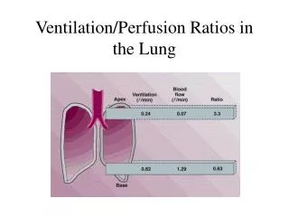
PHYSIOLOGY Ventilation & perfusion distribution Preferentially to non-dependent areas of lung V/Q ratio best at the base in upright position Non- symmetrical branching nature of airways & pulmonary vasculature accounts for 75% of ventilation perfusion distribution Gravity (25%) Galvin L ,GB Drummond, M.. Nirmalan. Distribution of blood flow and ventilation in the lung :- gravity is not the only factor . British J. Anaesth. 2007 ; 98: 420-428
LATERAL DECUBITUS - ADULT Dependent lung - Better ventilation & perfusion Hydrostatic pressure gradient Diaphragmatic contractility proportional to resting length of muscle . Greater contractility = Greater ventilation Resting length - Pressure of abdominal contents Oxygen consumption 3ml/kg/min
LATERAL DECUBITUS - INFANT Non –dependent lung better ventilation Chest floppy & compressible Dependent lung compressed FRC closer to residual volume Diaphragmatic contractility less significant Reduced hydrostatic pressure gradient Oxygen consumption 6-8 ml/kg/min
THORACIC SURGERY V/Q MISMATCH OCCURS - General anesthesia - Neuromuscular block - Mechanical ventilation - Single lung ventilation - Surgical retraction of lung - HPV (due to inhalational agents/vasodilators)
INDICATIONS -SLV ISOLATION / PROTECTION OF LUNG 1. Hemorrhage 2. Infective secretions – Pus 3. Pulmonary lavage COLLAPSE OF A LUNG – BETTER VISUALIZATION 1. Video Assisted Thoracoscopic Surgery (VATS) Pneumonectomy, Lobectomy 2. Intrathoracic non – pulmonary surgeries a. Vascular – PDA, Coarctation of aorta b. Vertebral – Anterior spinal fusion c. Esophageal – Hiatal hernia, Tracheo esophageal fistula d. Tumors – Mediastinal mass DIFFERENTIAL LUNG VENTILATION 1. Bronchopleural fistula 2. Unilateral lung cyst
CONTRAINDICATIONS -SLV Inability to place a device Severe ventilation perfusion mismatch
TECHNIQUES Double Lumen Tubes – Right & Left sided Univent Tubes Endobronchial Blockers Endotracheal Tubes
DOUBLE LUMEN TUBES (DLT) Two tubes (unequal length) molded together Shorter tracheal & longer bronchial tubes Two curves at almost 90⁰ - proximal & distal Cross section of tube is D shaped RIGHT LEFT
DOUBLE LUMEN TUBES • Two cuffs -Tracheal (proximal) & Bronchial (distal) • High volume low pressure cuffs –Don’t over inflate • Endobronchial cuff – Blue ,low volume & pressure • Rt.endobronchial cuff slot (Rt. upper lobe bronchus) • Radiographic markers near cuffs and slot CONNECTOR Bronchial cuff Site for clamp Radiographic markers Lumen for right upper lobe Fiberoptic scope or Suction Tracheal cuff
DOUBLE LUMEN TUBES Inserted under direct laryngoscopic view Stylet used Check cuffs before insertion Insert with distal curve concave anteriorly Remove stylet when tip is beyond larynx Rotate 900 to the bronchial side Proximal curve concave anteriorly (final position) Position confirmation – 1.Clinical - auscultation 2. Fiber optic scope
DOUBLE LUMEN TUBES Available sizes -26, 28, 32, 35, 37, 39 & 41Fr Chest X-Ray & CT Scan – Measure the bronchial diameter for DLT selection Proper size - deflated bronchial part <1-2mm of main stem bronchial lumen 8yr old (26Fr ) & 10 yr old (28Fr) Size in Fr = 4 x ID of endotracheal tube + 2 Proper size and length fiberoptic scope Left sided easier to place
DLT - ADVANTAGES Rapidity & ease of insertion Suctioning of individual lung Complete separation of lungs Ventilation of individual lung CPAP or Oxygen to the collapsed lung Placement possible in absence of fiberoptic scope Easy to convert from 2 lungs to SLV
DLT - DISADVANTAGES Cannot be used in less than 8 yr old Selection of size - difficult Difficult to place in abnormal airway Potential for airway trauma Require conversion to single lumen post op.
MARRARO PAEDIATRIC BILUMEN TUBE Two uncuffed tubes attached laterally PVC tubes with radiopaque line (Portex -U.K) Shorter tracheal tube attached to whole length of bronchial tube except a short part of the beginning Longer bronchial tube –Murphy eye. Bent at 50 angle before the eye Tubes end in lip shape facing outwards No spur for anchoring Bronchial lumen larger –different caliber tubes
MARRARO BILUMEN TUBE SINGLE CONNECTOR TRACHEAL INDIVIDUAL CONNECTORS BRONCHIAL RADIO OPAQUE LINE Photographs – Courtesy of Dr. Marraro
MARRARO BILUMEN TUBE G.Marraro. Selective endobronchial intubation in paediatrics: the Marraro Paediatric Bilumen Tube .Paed Anaesth 1994;4: 255-258
MARRARO BILUMEN TUBE Placed under direct laryngoscopic view Rotate to required bronchus after both tubes are below the cords Confirmation of correct position 1. Auscultation 2. Chest X – Ray 3. Fiber optic scope -1.8mm Right bronchial intubation easier Left bronchial intubation preferred Bronchial lumen larger –better fit & reduces leak Tube position at level of cords - one above the other O O
MARRARO BILUMEN TUBE Useful in children less than 3 years of age Humidification and warming of gases Increased resistance to air flow –small calibers Suction catheters marked –not to go beyond length Recommended PEEP < 10 cm H2O for tracheal < 15 cm H2O for bronchial External diameter = sum of internal diameters of both tubes + 0.4mm. (2.5/3 caliber tube =5.9 ext. diameter) Personal Communication –Dr. Marraro
MARRARO BILUMEN TUBE ADVANTAGES All the advantages of a double lumen tube in < 3yrs age Fiberoptic scope is not needed to confirm position Delivery of drugs (surfactant) to specific areas of lung DISADVANTAGES Experience & training required Dislodgement / obstruction of the tubes Trauma to larynx, trachea and bronchus Size chosen is empirical & arbitrary
UNIVENT TUBES Single lumen tube with an enclosed blocker Endobronchial blocker –separate channel Blocker is long & can be locked in position Blocks right / left bronchus Fiberoptic scope required for placement Pediatric sizes – ID 3.5 mm uncuffed 4.5 mm cuffed Lumen in blocker (not in 3.5mm) Pediatric age 6 years and above
UNIVENT TUBE –ADULT SIZE PROVIDE OXYGEN / SUCTION BLOCKER TO LOCK BLOCKER TO ATTACH BLOCKER IN PLACE
UNIVENT TUBES ADVANTAGES Displacement less since it is attached to the tube No need to change tube – postoperative period DISADVANTAGES Low volume high pressure cuff High resistance to gas flow Cannot use in < 6 years of age No suction, oxygen or CPAP to collapsed lung Lung collapse slow
ENDOBRONCHIAL BLOCKERS -IDEAL Shape which would stabilize it in the bronchus High volume/ low pressure at inflation Flexible & easy to manipulate Channel for deflation & suction Used external /internal to an endotracheal tube Available in adult & pediatric sizes
ENDOBRONCHIAL BLOCKERS - TYPES Fogarty catheters –vascular embolectomy catheters Balloon wedge catheters –Swan Ganz catheters Atrioseptostomy catheters Arndt Cook bronchial blockers – specifically designed for bronchi 4 Fr FOGARTY CATHETER BLIND END
FOGARTY CATHETERS Embolectomy catheter Placed within /outside of endotracheal tube Endobronchial intubation - for placement FOB – reposition & confirmation of placement Fluoroscopy –placement confirmation Sizes (Pediatric ) -3Fr, 4 Fr, 5 Fr Potential problem –dislodgement & difficulty in maintaining position Stylet removed & inflation maintained using a stopcock Tan GM, Tan- Kendrick AP. Bronchial diameter in children – use of the Fogarty catheter for lung isolation in children. Anaesth Intensive Care 2002; 30:615 -618
ARNDT COOK BRONCHIAL BLOCKERS Specifically designed for bronchus Sizes 5Fr, 7Fr & 9Fr (adult) Spherical / elliptical (9Fr) balloon Used in children 2 yrs and above Multiport adapter & FOB -Placement Adjustable guide loop (nylon ) Central channel -Nylon wire
ARNDT COOK BRONCHIAL BLOCKERS MULTI-PORT ADAPTER CIRCUIT 5Fr Arndt Cook Bronchial Blocker Nylon loop for scope Spherical & Elliptical Balloons of Blocker ( Cookmedical.com )
5Fr ARDNT COOK BLOCKER Most commonly used in children Placed outside the ETT in children <2 yrs ETT 2.5 - 4 mm –blocker outside Guide wire loop left in place for repositioning Max diameter of blocker = 2.5mm Central lumen diameter = 0.7 mm Balloon length = 1 cm& capacity = 1-3 ml Placement confirmed –FOB, auscultation & fluoroscopy
ADVANTAGES Able to block Rt. or Lt. bronchus Ventilation possible during placement Used with an endotracheal tube Connecter which locks blocker in place Suction possible CPAP possible Ease of removal Ease of 2 lung ventilation from single lung ventilation
DISADVANTAGES Dislodgement Frequent repositioning Non optimal rt. lung isolation Collapse of lung slower with smaller blockers Guide wire required for proper placement Pediatric FOB required Airway injury Bronchoscopy of isolated lung - impossible
INSERTION TECHNIQUES Multiport adapter and pediatric fiber optic scope Blocker guided into bronchus by endobronchial intubation, endotracheal tube removed and reinserted by the side of blocker Lianne L. Stepnenson, Christian Seefelder : Routine Extraluminal Use of the 5 Fr Arndt Endobronchial Blocker for One-Lung Ventilation in Children up to 24 Months of Age. J Cardiothorac Vasc Anesth 2010; 20 July On line. Ho AMH, Karmakar MK, Critchley LAH, et al: Placing the tip of the endotracheal tube at the carina and passing the endobronchial blocker through the Murphy eye may reduce the risk of blocker retrograde dislodgement during one-lung anaesthesia in small children. Br J Anaesth 2008;101: 690-693 Blocker inside ETT + /- Through Murphy eye Blocker outside ETT + /- Through Murphy eye
STANDARD ENDOTRACHEAL TUBE Advancing the endotracheal tube into the main stem bronchus opposite to site of surgery Collapse of lung in surgical area is by absorption atelectasis To advance tube into left bronchus – Turn head to right
DISADVANTAGES Inadequate seal of the bronchus Inadequate lung collapse on the operative side Failure of complete protection of ventilated lung from contaminants Inability to suction the non ventilated lung Inability to provide oxygen / CPAP to non ventilated lung Hypoxemia –blockade of upper lobe bronchus
SELECTION FOR A PATIENT Age of the patient Size of the patient Anatomy of the airway Skill & experience of the provider Availability of equipment Type of surgery
SLV – TRACHEOSTOMY DEPENDING ON AGE / SIZE OF TRACHEOSTOMY 1. Endobronchial blocker - Fogarty / Arndt Cook (co-axially or along the tracheostomy tube ) 2. Single lumen endotracheal tube directed to main stem bronchus via the stoma
TIPS - AVOIDING HYPOXEMIA Single lung ventilation only when needed PEEP to the dependent lung Oxygen & CPAP to the collapsed lung Increasing Fi O2 Check the tube Treat any hemodynamic instability Maintain CO2 within normal limits Clamp pulmonary artery (Pneumonectomy)
SUMMARY Knowledge of patient’s airway anatomy Physiology of single lung ventilation Details of surgical procedure Proper selection Check all equipments before use Recruit help if needed
REFERENCES Andrew B. Lumb: Nunn’s Applied Respiratory Physiology 6th edition Elsevier Butterworth Heinemann Pg 110-113 & pg 297-326 Remolina C, Khan AU, Santiago TV, Edelman NH: Positional hypoxemia in unilateral lung disease. N Engl J Med 1981;304: 523-5. Heaf DP, Helms P, Gordon I, Turner HM: Postural effects on gas exchange in infants. N Engl J Med 1983;308: 1505-8. Gregory B. Hammer: Pediatric Thoracic Anaesthesia. Anesth Analg 2001; 92: 1449-64. Gregory B.Hammer ,Brett G. Fitzmaurice, Jay B. Brodsky: Methods for Single-Lung Ventilation in Pediatric Patients. Anesth Analg 1999; 89: 1426-9 . Oliver Bagshaw, Steven Cray. Anesthesia for Thoracic Surgery. In: Dakshesh H. Parikh, David C.G. Crabbe, Alexander W. Auldist, Steven S. Rothenberg. Editors, Pediatric Thoracic Surgery , Springer. London 2009 : 57-74 Dinesh K. Choudhry: Single-Lung Ventilation in Pediatric Anesthesia. Anesthesiology Clin N America 2005;23:693-708