Download
1 / 59
590 likes | 782 Vues
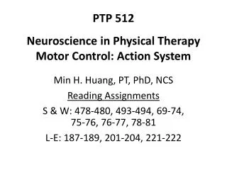
PTP 512 Neuroscience in Physical Therapy Motor Control: Action System. Min H. Huang, PT, PhD, NCS Reading Assignments S & W: 478-480, 493-494, 69-74, 75-76, 76-77, 78-81 L-E: 187-189, 201-204, 221-222. Objectives. Describe and demonstrate spinal modulated reflexes

E N D
PTP 512Neuroscience in Physical TherapyMotor Control: Action System Min H. Huang, PT, PhD, NCS Reading Assignments S & W: 478-480, 493-494, 69-74, 75-76, 76-77, 78-81 L-E: 187-189, 201-204, 221-222
Objectives • Describe and demonstrate spinal modulated reflexes • Differentiate reflexes vs. voluntary movement • Differentiate feedback vs. feedforward control • Explain recruitment patterns of motor units to generate force in muscles • Indicate the role of each neural structure involved in motor control • Describe the clinical implications following lesions to each neural structure
Movement Control Reflexic Voluntary With conscious intention to move • Stereotyped motor response to a specific sensory stimulus • Neurons involved • Sensory neurons • Interneurons • Motor neurons • Reflex responses are often complex and can change depending on the task
Rapid wrist flexion movement in a normal subject Berardelli, 1996
Rapid flexion movements at the elbow Parkinson’s Disease The three traces in each panel are, from top to bottom, rectified biceps, and triceps EMG, angular position. Cerebellar deficits Berardelli, 1996
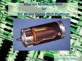
Motor Neurons • Alpha motor neuron (lower motor neuron) • Motor neuron pool • Motor unit • Motor unit innervation ratio (MUIR) • Compare MUIR between finger extensor vs. deltoid muscle • A motor neuron controls the amount of force that is exerted by muscle fibers Click to view animationFigure 1.4. Byrne, 1997. http://nba.uth.tmc.edu/neuroscience/s3/chapter01.html
Control of Muscle Force: Rate Cod • ↑in the rate of action potentials fired by the motor neuron causes ↑in the amount of force that the motor unit generates. Click to view animation. Figure 1.5. Byrne, 1997. http://nba.uth.tmc.edu/neuroscience/s3/chapter01.html
Control of Muscle Force: Size Principle • When the input to motor neurons increases, smaller motor neurons are recruited and fired action potentials first. • Small motor neurons innervate slow-twitch fibers; Intermediate-sized motor neurons innervate fast-twitch, fatigue-resistant fibers; Large motor neurons innervate fast-twitch, fatigable muscle fibers. Click to view animation. Figure 1.6. Byrne, 1997. http://nba.uth.tmc.edu/neuroscience/s3/chapter01.html
Stretch Reflex (DTR) Stimulus excites muscle spindle which sends signals via Ia afferents to α motor neuron • Excitation of agonist • Excitation of synergist • Inhibition of antagonist (i.e. reciprocal inhibition or reciprocal innervation) • Click to view animation Shunway-Cook, 2007
Stretch Reflex (DTR) Muscle spindle information also goes to supraspinal regions via a long-loop reflex pathway, i.e. transcortical reflex, or functional stretch reflex. Click to view abnormal DTR Shunway-Cook, 2007
Task-Dependent Modulation of Stretch Reflexes Pearson, 2008 For example, perturbation of one arm causes different responses in the contralateral arm, depending on the task.
Task instructions and environmental stiffness modify stretch reflex PTB: rapid stretches to biceps SLR: short-latency stretch reflex (monosynaptic) LLR: long-latency stretch reflex (polysynaptic and transcortical) Shemmell, 2009
Flexor Withdrawal Noxious stimulus to skin causes protective withdrawal of limb through • Excitation of ipsilateral flexor muscles • Inhibition of ipsilateral antagonistic extensor muscles Pearson, 2008 • Click to view animation
Crossed Extension Typically occurs in conjunction with flexor withdrawal. Noxious stimulus causes contralateral • Excitation of extensor muscles • Inhibition of flexor muscles Pearson, 2008 • Click to view animation
Autogenic Inhibition Contraction or stretch of muscle pulls on the Golgi tendon organ (GTO). GTO sends signal to α motor neuron via Ib afferents and causes • Inhibition of agonist muscle • Excitation of antagonist muscle Shunway-Cook, 2007 • Click to view animation
Autogenic Inhibition: GTO Function • Protect muscle if contracting too hard, or fatigue • Clasp-knife reflex thought to be mediated in part through autogenic inhibition • Related to GTO Young, 2008
Plantar and Finger Flexors Reflexes Plantar (Babinski) Finger flexor (Hoffman) Tap or flick the terminal phalanx of the 3rd or 4th digit Positive Response flexion of terminal phalanx of thumb UE equivalent of Babinski • Stroke foot firmly • Normal response • toes plantarflex • Positive response • great toe flexes toward the top of the foot and some spraying of other toes
Babinski Sign • Positive Babinski or Hoffman reflexes are signs of upper motor neuron lesions Normal response Positive response View Hoffman Reflex http://neuroexam.com/neuroexam/content.php?p=33 View Babinski Reflex http://neuroexam.com/neuroexam/content.php?p=32
DESIRED OUTPUT EFFECTOR OUTPUT COMPARATOR – + EFFECTOR SENSOR
Feedback Control Shunway-Cook, 2007
ADVANCED INFORMAITON SENSOR DESIRED OUTPUT EFFECTOR OUTPUT
Feedforward Control Shunway-Cook, 2007
Feedback vs. Feedforward Control • Feedforward control uses initial inputs from vision • Feedback control uses somatosensory inputs from arm Shunway-Cook, 2007
Prefrontal Cortex • Strategic planning, decision to move • Motivation • Changing strategy to move, adaptive • Selection of appropriate actions for a particular behavioral context
Posterior Parietal Cortex • Ensuring that movements are targeted accurately to objects in external space • Processing spatial relationships of objects in the world • Building a representation of external space
Primary Motor Cortex (Area 4): Execution of Voluntary Movement Evarts’ study (1968) monkeys made wrist flexion/extension • Fire 5-100 msec before movement onset • Encodes force of movement: the rate of neuronal firing ↑ with increased resistance to movement View animationFigure 3.7 http://neuroscience.uth.tmc.edu/s3/chapter03.html
Primary Motor Cortex (Area 4): Execution of Voluntary Movement • Encodes direction anddistanceof movement Vectors show firing of neurons to movements in different directions ( indicates max response) Monkeys moved lever toward light
Primary Motor Cortex (Area 4): Execution of Voluntary Movement • Encode the speedof movement • Movement velocity profile is typically “bell-shaped” • Firing rates of neurons correlate with acceleration and deceleration of the velocity profile Hand velocity during a reaching task. Rosenbaum, 1995
True or false: “All of the neurons that control the biceps muscle may be located together” Incorrect! Because • Primary Motor Cortex (M1) does not generally control individual muscles directly, but appears to control movements of individual body parts or sequences of movements • Stimulation of small regions of MI causes movements that require activity of many muscle. Conversely, movements of individual muscles are correlated with activity of widespread areas of the MI.
Pre-motor Cortex • Involved in more complex, task-oriented process of movement control than primary motor cortex, and the selection of appropriate motor plans for voluntary movements. View animation Figure 3.2 http://neuroscience.uth.tmc.edu/s3/chapter03.html Lundy-Ekman, 2007
Premotor Cortex • Signal the preparation for movement: “motor set neurons” fire when preparing to make a movement, and then turn silent during movement execution • Signal various sensory aspects associated with particular motor acts: “mirror neurons” fire selectively during a particular action, e.g. drinking coffee, and also when seeing or hearing others drinking coffee. View animation Figure 3.10 http://neuroscience.uth.tmc.edu/s3/chapter03.htm
Videos on mirror neurons Mirror Neuron. NOVA Science Now. http://www.pbs.org/wgbh/nova/body/mirror-neurons.html Mirror Neuron. Human Spark. http://www.pbs.org/wnet/humanspark/video/web-exclusive-video-mirror-neurons/404/
Premotor Cortex • Sensitive to the behavioral context of a particular movement: neurons fire selectively to the inferred intentions of a movement, not just the movement itself • Signals correct and incorrect actions • Involved in voluntary movements activated by external stimuli, e.g. sensory stimuli, and visually guided sequences of movement View animation Figure 3.12 http://neuroscience.uth.tmc.edu/s3/chapter03.html
Supplemental Motor Area (SMA) • Involved in the selection of complex sequential movements and the coordination of bilateral movements • Bilateral SMA are activated during the execution of complex sequential movements, and also during mental rehearsal of complex sequential movements • Mental rehearsal is a treatment technique involving the SMA
Shumway-Cook, 2007 View animation Figure 3.13. http://neuroscience.uth.tmc.edu/s3/chapter03.html
Supplemental Motor Area (SMA) • Involved in learning sequential movements • Involved in selecting movements that are initiated internally • Transform kinematic information (position, velocity) of a movement plan to dynamic (force) information of a movement plan
Cerebellum • Review Lundy’s Fig 10.16
Fine tuning of movement in feedback control: cerebellum compare “efferent copy” of motor program with incoming sensory feedback to correct for deviations from the intended movement outcome.
Motor learning and feedforward control: cerebellum evaluates sensory information and movement errors to modify the strength of synaptic connections in cerebellar cortex. As a result, future movements in a similar context will be modified.
Cerebellum • Non-motor function • Timing in perceptual tasks: may serve as a centralized clock for body functions • Recall of habits/skills learned through repetition • Language process related to movement, e.g. verbs
Cerebellum: Clinical Dysfunction http://library.med.utah.edu/neurologicexam/html/coordination_abnormal.html#12 • Speech Rapid Alternating Movements • Dysarthria • Tremor • Hand Rapid Alternating Movements • Finger-to-nose • Foot rapid alternating movements • Tandem Gait The University of Utah 2001
Cerebellum: Clinical Dysfunction • Muscle tone ↓ • Ataxic gait: unable to walk heel-to-toe • Truncal ataxia: involving trunk • Appendicular ataxia: involving limbs • Dysarthria: slurred, uncoordinated speech • Nystagmus: visual guidance of movement deteriorates
Cerebellum: Clinical Dysfunction • Dysmetria • Hypermetria is most common: past-pointing, overshooting of target • Hypometria • Intention tremor • Dysdiadochokinesis • Impaired sequential movements • Test: rapid alternating movements (RAM)