Download
1 / 19
220 likes | 544 Vues
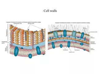
In most plant tissues The cell walls and cytosol are continuous from cell to cell The cytoplasmic continuum Is called the symplast The apoplast Is the continuum of cell walls plus extracellular spaces. Key. Symplast. Apoplast. Transmembrane route. Apoplast. The symplast is the

E N D
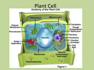
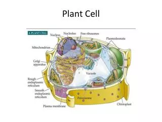
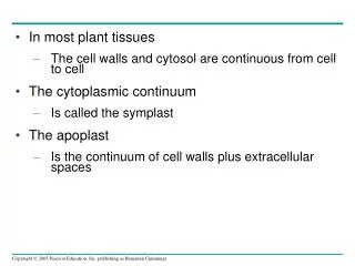
In most plant tissues • The cell walls and cytosol are continuous from cell to cell • The cytoplasmic continuum • Is called the symplast • The apoplast • Is the continuum of cell walls plus extracellular spaces
Key Symplast Apoplast Transmembrane route Apoplast The symplast is the continuum of cytosol connected by plasmodesmata. The apoplast is the continuum of cell walls and extracellular spaces. Symplast Symplastic route Apoplastic route (b) Transport routes between cells. At the tissue level, there are three passages: the transmembrane, symplastic, and apoplastic routes. Substances may transfer from one route to another. Figure 36.8b
Functions of the Symplast and Apoplast in Transport • Water and minerals can travel through a plant by one of three routes • Out of one cell, across a cell wall, and into another cell • Via the symplast • Along the apoplast
Casparian strip Endodermal cell Pathway along apoplast Pathway through symplast 1 Uptake of soil solution by the hydrophilic walls of root hairs provides access to the apoplast. Water and minerals can then soak into the cortex along this matrix of walls. Casparian strip 2 Plasma membrane 1 Minerals and water that cross the plasma membranes of root hairs enter the symplast. Apoplastic route 2 Vessels (xylem) 3 As soil solution moves along the apoplast, some water and minerals are transported into the protoplasts of cells of the epidermis and cortex and then move inward via the symplast. Root hair Symplastic route Epidermis Endodermis Vascular cylinder Cortex 5 4 Endodermal cells and also parenchyma cells within the vascular cylinder discharge water and minerals into their walls (apoplast). The xylem vessels transport the water and minerals upward into the shoot system. Within the transverse and radial walls of each endodermal cell is the Casparian strip, a belt of waxy material (purple band) that blocks the passage of water and dissolved minerals. Only minerals already in the symplast or entering that pathway by crossing the plasma membrane of an endodermal cell can detour around the Casparian strip and pass into the vascular cylinder. Figure 36.9 • Lateral transport of minerals and water in roots
The Roles of Root Hairs, Mycorrhizae, and Cortical Cells • Much of the absorption of water and minerals occurs near root tips, where the epidermis is permeable to water and where root hairs are located • Root hairs account for much of the surface area of roots
2.5 mm • Most plants form mutually beneficial relationships with fungi, which facilitate the absorption of water and minerals from the soil • Roots and fungi form mycorrhizae, symbiotic structures consisting of plant roots united with fungal hyphae Figure 36.10
Concept 36.3: Water and minerals ascend from roots to shoots through the xylem • Plants lose an enormous amount of water through transpiration, the loss of water vapor from leaves and other aerial parts of the plant • The transpired water must be replaced by water transported up from the roots
Pushing Xylem Sap: Root Pressure • At night, when transpiration is very low • Root cells continue pumping mineral ions into the xylem of the vascular cylinder, lowering the water potential • Water flows in from the root cortex • Generating root pressure
Cohesion and Adhesion in the Ascent of Xylem Sap • The transpirational pull on xylem sap • Is transmitted all the way from the leaves to the root tips and even into the soil solution • Is facilitated by cohesion and adhesion
Xylem sap Outside air Y = –100.0 MPa Mesophyll cells Stoma Water molecule Leaf Y (air spaces) = –7.0 MPa Transpiration Atmosphere Leaf Y (cell walls) = –1.0 MPa Xylem cells Adhesion Cell wall Water potential gradient Trunk xylem Y = – 0.8 MPa Cohesion, by hydrogen bonding Cohesion and adhesion in the xylem Water molecule Root xylem Y = – 0.6 MPa Root hair Soil Y = – 0.3 MPa Soil particle Water Water uptake from soil Figure 36.13 • Ascent of xylem sap
Xylem Sap Ascent by Bulk Flow: A Review • The movement of xylem sap against gravity • Is maintained by the transpiration-cohesion-tension mechanism
Concept 36.4: Stomata help regulate the rate of transpiration • Leaves generally have broad surface areas • And high surface-to-volume ratios
20 µm Figure 36.14 • Both of these characteristics • Increase photosynthesis • Increase water loss through stomata
Cells turgid/Stoma open Cells flaccid/Stoma closed (a) Changes in guard cell shape and stomatal opening and closing (surface view). Guard cells of a typical angiosperm are illustrated in their turgid (stoma open) and flaccid (stoma closed) states. The pair of guard cells buckle outward when turgid. Cellulose microfibrils in the walls resist stretching and compression in the direction parallel to the microfibrils. Thus, the radial orientation of the microfibrils causes the cells to increase in length more than width when turgor increases. The two guard cells are attached at their tips, so the increase in length causes buckling. Radially oriented cellulose microfibrils Cell wall Vacuole Guard cell Figure 36.15a • Each stoma is flanked by guard cells • Which control the diameter of the stoma by changing shape
H2O H2O H2O H2O (b) Role of potassium in stomatal opening and closing. The transport of K+ (potassium ions, symbolized here as red dots) across the plasma membrane and vacuolar membrane causes the turgor changes of guard cells. H2O K+ H2O H2O H2O H2O H2O Figure 36.15b • Changes in turgor pressure that open and close stomata • Result primarily from the reversible uptake and loss of potassium ions by the guard cells
Movement from Sugar Sources to Sugar Sinks • Phloem sap • Is an aqueous solution that is mostly sucrose • Travels from a sugar source to a sugar sink
A sugar source • Is a plant organ that is a net producer of sugar, such as mature leaves • A sugar sink • Is an organ that is a net consumer or storer of sugar, such as a tuber or bulb
Sieve-tube member Companion (transfer) cell Mesophyll cell Cell walls (apoplast) Plasma membrane Plasmodesmata (a) Sucrose manufactured in mesophyll cells can travel via the symplast (blue arrows) to sieve-tube members. In some species, sucrose exits the symplast (red arrow) near sieve tubes and is actively accumulated from the apoplast by sieve-tube members and their companion cells. Phloem parenchyma cell Bundle- sheath cell Figure 36.17a Mesophyll cell • Sugar must be loaded into sieve-tube members before being exposed to sinks • In many plant species, sugar moves by symplastic and apoplastic pathways
High H+ concentration Cotransporter H+ Proton pump S (b) A chemiosmotic mechanism is responsible for the active transport of sucrose into companion cells and sieve-tube members. Proton pumps generate an H+ gradient, which drives sucrose accumulation with the help of a cotransport protein that couples sucrose transport to the diffusion of H+ back into the cell. Key ATP Sucrose H+ H+ Apoplast S Low H+ concentration Symplast Figure 36.17b • In many plants • Phloem loading requires active transport • Proton pumping and cotransport of sucrose and H+ • Enable the cells to accumulate sucrose