Prolapse and Incontinence
210 likes | 495 Vues
Prolapse and Incontinence. Craig Dyson Sioned Griffiths October 2013. Contents. Normal Anatomy Causes of prolapse Types of Prolapse Investigation Management. Anatomy. Anatomy. Anatomy. Prolapse. “To fall out”

Prolapse and Incontinence
E N D
Presentation Transcript
Prolapse and Incontinence Craig Dyson Sioned Griffiths October 2013
Contents • Normal Anatomy • Causes of prolapse • Types of Prolapse • Investigation • Management
Prolapse • “To fall out” • Protrusion of an organ or structure beyond its normal confines and with an epithelial surface • Genitourinary prolapse – Descent of one or more of pelvic organs. • 41% of 50-79 year old’s but uncertain • Uterocoele, Cystocoele, Rectocoele, Enterocoele
Pathophysiology • Levator Ani/Endopelvic Fascia important • Damage to these structures can occur through: • Trauma • Neuropathic Injury • Disruption/Stretching • Multifactorial – Orientation of bones may be a factor.
Risk Factors • Increasing Age (Double risk with every decade) • Vaginal Delivery • Increasing parity • Obesity • Spina Bifida • Pregnancy Variables • Macrosomia • Prolonged 2nd stage • Episiotomy • Use of forceps/oxytocin • FH of prolapse • Constipation • Connective Tissue Disorder • Occupation
Types • Anterior • Urethrocoele • Urinary Stress Incontinence • Rare • Cystocoele • Increased frequency • UTI • Sensation of mass • No Symptoms • Both • Most Common
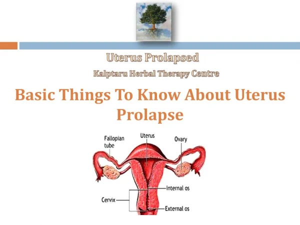
Types • Middle • Uterine Prolapse • Vaginal Vault Prolapse • Post Hysterectomy • Assoc with cystocoele, rectocoele and enterocoele. • Retention • Enterocoele • Pouch of Douglas • Cough Impulse
Types • Posterior • Rectocoele
POPQ System • Pelvic Organ Prolapse Quantification System • Valsalva - ? Left Lateral • Stage 0 • Stage 1 – 1cm above hymen • Stage 2 - Within 1 cm of hymen • Stage 3 - >1cm below plane of hymen but <2cm of total length of vagina • Stage 4 – Complete eversion of vagina
Symptoms • General • Fullness • Sensation of bulge • Backache • Urinary • Incontinence • Frequency • Coital • Dypareunia • Flatus • Bowel • Constipation/Incontinence • Need to apply digital pressure
Investigations • History and Examination • Urinalysis • Post-Voidal Urine volume testing • Urodynamics • US • Urea/Creatinine
Management • Conservative • Watchful Waiting • Lifestyle Modification • Pelvic Floor Exercises • Evidence? • Vaginal Oestrogen Creams • Pessary
Pessary • Inserted into vagina to reduce prolapse • Made of silicon or plastic or Soaked in wine… • Good short term option
Management • Surgical • Effective • Re-operation required in 29% of cases • Fitness of patient • Sexually Active • Surgeons Advice
Surgery • Anterior Colporrhaphy • Involves plication of anterior vaginal wall to reinforce. • Hysterectomy • Sacrospinous Fixation • Unilateral or bilateral fixation of uterus to sacrospinous ligament • Sacocolpoplexy • Mesh used to attach top of vagina to sacrum.
Summary • Prolapse is increasingly common with age. • Can be classified according to compartment or level of prolapse • Can be clear on examination • Good conservative and surgical options available • Good prognosis
References • Pessary treatment for pelvic organ prolapse and health-related quality of life: a review. Lamers BH, Broekman BM, Milani AL - Int Urogynecol J (2011) • Rev Urol. 2004; 6(Suppl 5): S2–S10. PMCID: PMC1472875. Female Pelvic Floor Anatomy: The Pelvic Floor, Supporting Structures, and Pelvic Organs. Sender Herschorn • Herschorn S, Carr LK. In: Campbell’s Urology. 2002:1092–1139. • Rectocele | Vaginal Surgery & Urogynecology Institute .vaginalsurgeryandurogynecologyinstitute.com • Int J Med Sci 2012; 9(10):894-900. doi:10.7150/ijms.4829. Three-dimensional Ultrasound Appearance of Pelvic Floor in Nulliparous Women and Pelvic Organ Prolapse Women. Tao Ying Corresponding address, Qin Li, Lian Xu, Feifei Liu, Bing Hu • http://www.patient.co.uk/health/Genitourinary-GU-Prolapse.htm • www.pelvicfloor.com/knowledge/imagelibrary/1/img/1.jpg • www.bristolsurgery.com/images/Preop%20Rectocele.jpg