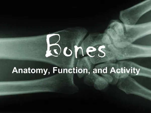
Bones
Bones. The Support System and More. Skeletal Cartilage. Contains no blood vessels or nerves Surrounded by the perichondrium (dense irregular connective tissue) that resists outward expansion Three types Hyaline Elastic fibrocartilage. Hyaline Cartilage.

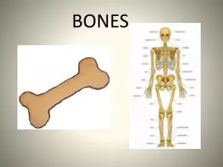
Bones
E N D
Presentation Transcript
Bones The Support System and More
Skeletal Cartilage • Contains no blood vessels or nerves • Surrounded by the perichondrium (dense irregular connective tissue) that resists outward expansion • Three types • Hyaline • Elastic • fibrocartilage
Hyaline Cartilage • the most abundant skeletal cartilage • Support, flexibility, and resilience • Present in these cartilages: • Articular – covers the ends of long bones • Costal – connects the ribs to the sternum • Respiratory – makes up the larynx and reinforces air passages • Nasal – supports the nose
Elastic Cartilage • Similar to hyaline cartilage but contains elastic fibers • Found in: • the external ear • the epiglottis
Fibrocartilage • Highly compressed with great tensile strength • Contains collagen fibers • Found in: • menisci of the knee • intervertebral discs
Growth of Cartilage • Appositional • cells in the perichondrium secrete matrix against the external face of existing cartilage • Interstitial • lacunae-bound chondrocytes inside the cartilage divide and secrete new matrix, expanding the cartilage from within • Calcification of cartilage occurs • During normal bone growth • During old age
Classification of Bones: By Shape • Long bones are longer than they are wide (e.g., humerus)
Classification of Bones: By Shape • Flat bones are thin, flattened, and a bit curved (e.g., sternum, and most skull bones)
Classification of Bones: By Shape • Irregular bones – bones with complicated shapes (e.g., vertebrae and hip bones)
Function of Bones • Support • form the framework that supports the body and cradles soft organs • Protection – provide a protective case for the brain, spinal cord, and vital organs • Movement – provide levers for muscles • Mineral storage – reservoir for minerals, especially calcium and phosphorus • Blood cell formation – hematopoiesis occurs within the marrow cavities of bones
Gross Anatomy of Bones: Bone Textures • Compact bone – dense outer layer • Spongy bone – honeycomb of trabeculae filled with yellow bone marrow
Structure of Long Bone Figure 6.3
Structure of Long Bone • Long bones consist of a diaphysis and an epiphysis • Diaphysis • Tubular shaft that forms the axis of long bones • Composed of compact bone that surrounds the medullary cavity • Yellow bone marrow (fat) is contained in the medullary cavity
Structure of Long Bone • Epiphyses • Expanded ends of long bones • Exterior is compact bone, and the interior is spongy bone • Joint surface is covered with articular (hyaline) cartilage • Epiphyseal line separates the diaphysis from the epiphyses
Structure of Long Bone Figure 6.3
Bone Membranes • Periosteum – double-layered protective membrane • Outer fibrous layer is dense regular connective tissue • Inner osteogenic layer is composed of osteoblasts and osteoclasts • Richly supplied with nerve fibers, blood, and lymphatic vessels, which enter the bone via nutrient foramina • Secured to underlying bone by Sharpey’s fibers • Endosteum – delicate membrane covering internal surfaces of bone
Structure of Short, Irregular, and Flat Bones • Thin plates of periosteum-covered compact bone on the outside with endosteum-covered spongy bone (diploë) on the inside • Have no diaphysis or epiphyses • Contain bone marrow between the trabeculae
Location of Hematopoietic Tissue (Red Marrow) • In infants • Found in the medullary cavity and all areas of spongy bone • In adults • Found in the diploë of flat bones, and the head of the femur and humerus
Cells of Bone • Osteoblasts – bone-forming cells • Osteocytes – mature bone cells • Osteoclasts – large cells that resorb or break down bone matrix
Chemical Composition of Bone: Organic • Osteoid – unmineralized bone matrix composed of proteoglycans, glycoproteins, and collagen
Chemical Composition of Bone: Inorganic • Hydroxyapatites, or mineral salts • Sixty-five percent of bone by mass • Mainly calcium phosphates • Responsible for bone hardness and its resistance to compression
Developmental Aspects of Bones • Mesoderm gives rise to embryonic mesenchymal cells, which produce membranes and cartilages that form the embryonic skeleton • The embryonic skeleton ossifies in a predictable timetable that allows fetal age to be easily determined from sonograms • At birth, most long bones are well ossified (except for their epiphyses)
Developmental Aspects of Bones • By age 25, nearly all bones are completely ossified • In old age, bone resorption predominates • A single gene that codes for vitamin D docking determines both the tendency to accumulate bone mass early in life, and the risk for osteoporosis later in life
Formation of Bone • Intramembranous ossification – bone develops from a fibrous membrane • Formation of most of the flat bones of the skull and the clavicles • Endochondral ossification – bone forms by replacing hyaline cartilage • Uses hyaline cartilage “bones” as models for bone construction • Requires breakdown of hyaline cartilage prior to ossification
Stages of Intramembranous Ossification Figure 6.7.1
Stages of Intramembranous Ossification Figure 6.7.2
Stages of Intramembranous Ossification Figure 6.7.3
Stages of Intramembranous Ossification Figure 6.7.4
Endochondral Ossification • Begins in the second month of development • Uses hyaline cartilage “bones” as models for bone construction • Requires breakdown of hyaline cartilage prior to ossification
Stages of Endochondral Ossification Secondary ossification center Articular cartilage Epiphyseal blood vessel Spongy bone Deteriorating cartilage matrix Hyaline cartilage Epiphyseal plate cartilage Spongy bone formation Primary ossification center Medullarycavity Bone collar Blood vessel of periosteal bud 1 2 Formation of bone collar around hyaline cartilage model. Cavitation of the hyaline cartilage within the cartilage model. 3 4 Invasion of internal cavities by the periosteal bud and spongy bone formation. Formation of the medullary cavity as ossification continues; appearance of secondary ossification centers in the epiphyses in preparation for stage 5. 5 Ossification of the epiphyses; when completed, hyaline cartilage remains only in the epiphyseal plates and articular cartilages
Long Bone Growth and Remodeling Figure 6.10
Functional Zones in Long Bone Growth • Growth zone – cartilage cells undergo mitosis, pushing the epiphysis away from the diaphysis • Transformation zone – older cells enlarge, the matrix becomes calcified, cartilage cells die, and the matrix begins to deteriorate • Osteogenic zone – new bone formation occurs
Long Bone Growth and Remodeling • Growth in length – cartilage continually grows and is replaced by bone as shown • Remodeling – bone is resorbed and added by appositional growth as shown in the next slide
Appositional Growth of Bone Centralcanal of osteon Periosteal ridge Penetrating canal Periosteum Artery Osteoblasts beneath the periosteum secrete bone matrix, forming ridges that follow the course of periosteal blood vessels. As the bony ridges enlarge and meet, the groove containing the blood vessel becomes a tunnel. 1 The periosteum lining the tunnel is transformed into an endosteum and the osteoblasts just deep to the tunnel endosteum secrete bone matrix, narrowing the canal. 2 As the osteoblasts beneath the endosteum form new lamellae, a new osteon is created. Meanwhile new circumferential lamellae are elaborated beneath the periosteum and the process is repeated, continuing to enlarge bone diameter. 3 4
Hormonal Regulation of Bone Growth During Youth • During infancy and childhood, epiphyseal plate activity is stimulated by growth hormone • During puberty, testosterone and estrogens: • Initially promote adolescent growth spurts • Cause masculinization and feminization of specific parts of the skeleton • Later induce epiphyseal plate closure, ending longitudinal bone growth
Bone Deposition • Occurs where bone is injured or added strength is needed • Requires a diet rich in protein, vitamins C, D, and A, calcium, phosphorus, magnesium, and manganese • Alkaline phosphatase is essential for mineralization of bone • Sites of new matrix deposition are revealed by the: • Osteoid seam – unmineralized band of bone matrix • Calcification front – abrupt transition zone between the osteoid seam and the older mineralized bone
Bone Resorption • Accomplished by osteoclasts • Resorption bays – grooves formed by osteoclasts as they break down bone matrix • Resorption involves osteoclast secretion of: • Lysosomal enzymes that digest organic matrix • Acids that convert calcium salts into soluble forms • Dissolved matrix is transcytosed across the osteoclast’s cell where it is secreted into the interstitial fluid and then into the blood
Importance of Ionic Calcium in the Body • Calcium is necessary for: • Transmission of nerve impulses • Muscle contraction • Blood coagulation • Secretion by glands and nerve cells • Cell division
Control of Remodeling • Two control loops regulate bone remodeling • Hormonal mechanism maintains calcium homeostasis in the blood • Mechanical and gravitational forces acting on the skeleton
Hormonal Mechanism Figure 6.12
Response to Mechanical Stress • Wolff’s law – a bone grows or remodels in response to the forces or demands placed upon it • Observations supporting Wolff’s law include • Long bones are thickest midway along the shaft (where bending stress is greatest) • Curved bones are thickest where they are most likely to buckle
Response to Mechanical Stress • Trabeculae form along lines of stress • Large, bony projections occur where heavy, active muscles attach
Response to Mechanical Stress Figure 6.13
Bone Fractures (Breaks) • Classified by: • The position of the bone ends after fracture • The completeness of the break • The orientation of the bone to the long axis • Whether or not the bones ends penetrate the skin
Types of Bone Fractures • Nondisplaced – bone ends retain their normal position • Displaced – bone ends are out of normal alignment • Complete – bone is broken all the way through • Incomplete – bone is not broken all the way through • Linear – the fracture is parallel to the long axis of the bone
Types of Bone Fractures • Transverse – the fracture is perpendicular to the long axis of the bone • Compound (open) – bone ends penetrate the skin • Simple (closed) – bone ends do not penetrate the skin
Common Types of Fractures • Comminuted – bone fragments into three or more pieces; common in the elderly • Spiral – ragged break when bone is excessively twisted; common sports injury • Depressed – broken bone portion pressed inward; typical skull fracture • Compression – bone is crushed; common in porous bones
Common Types of Fractures • Epiphyseal – epiphysis separates from diaphysis along epiphyseal line; occurs where cartilage cells are dying • Greenstick – incomplete fracture where one side of the bone breaks and the other side bends; common in children