Endocrine hypertension
220 likes | 552 Vues
Endocrine hypertension. Except for: acromegaly, thyrotoxicosis, hypothyroidism, primary hyperparathyroidism. A few remarks on:. Primary hyperaldosteronism Congenital adrenal hyperplasia (due to: 17 -hydroxylase deficiency, 11-hydroxylase deficiency Cushing’s syndrome

Endocrine hypertension
E N D
Presentation Transcript
Endocrine hypertension Except for: acromegaly, thyrotoxicosis, hypothyroidism, primary hyperparathyroidism
A few remarks on: • Primary hyperaldosteronism • Congenital adrenal hyperplasia (due to: 17-hydroxylase deficiency, 11-hydroxylase deficiency • Cushing’s syndrome • Pheochromocytoma • Hypertension of renal origin
PRIMARY HYPERALDOSTERONISM • Sodium and fluid retention, expansion of ECFV and plasma volume, increased cardiac output • Vasoconstriction, increased total peripheral resistance Typical features: hypertension, hypokalemia, metabolic alkalosis, supression of the renin-angiotensin system ( PRA), Source of aldosterone:adenoma (75%), micro- or macronodular hyperplasia (idiopathic hyperaldosteronism) of zona glomerulosa K+ depletion impaired glucose tolerance, impaired urinary concentrating ability, postural hypotension Other hormones of zona glomerulosa: DOC (deoxycorticosterone), corticosterone, 18-OH-corticosterone
Clinical features Symptoms of hypokalemia: fatigue, loss of stamina, weakness, lassitude, increased thirst, polyuria, paresthesias, orthostatic hypotension Symptoms of alkalosis: a possitive Trousseau or Chvostek sign Hypertension No edema The most common cause of hypokalemia in hypertensive patients is diuretic therapy! A low Na+ diet, by reducing delivery of Na+ to aldosterone-sensitive sites in distal nephron, can reduce renal K+ secretion and thus correct hypokalemia. Average diet contains >120 mmol of Na+ per day Hormonal assessment: Plasma renin activity (PRA) Plasma aldosterone Urinary aldosterone excretion
Hormonal assessment Basal conditions: around 8 A.M. after at least 4 hrs. of recumbency (unrestricted salt diet): PRA, plasma aldosterone Stimulation test: a 4-hour upright posture furosemide i.v. ADENOMA: suppression of PRA, high basal plasma aldosterone level, no significant change or a frank decrease on stimulation. HYPERPLASIA: suppression of PRA, lower basal plasma aldosterone level (< 25 ng/dl), an increase on stimulation Saline infusion test (suppression): 2 L 0.9% NaCl over 2 hrs.: no suppression of plasma aldosterone
Location of adenoma CT or MRI imaging Adrenal scintigraphy:131I-iodocholesterol Adrenal vein catheterization: measurement and comparison of aldosterone levels Treatment Adenoma: unilateral adrenalectomy Hyperplasia: spironolactone Preoperative preparation: Spironolactone: 200-300 mg/d (4-6 weeks), maintenance dose 75-100 mg/d; reduces ECFV, promotes K+ retention, activates the suppressed renin-angiotensin system, prevents postoperative hypoaldosteronism. Side effects: rashes, gynecomastia, impotence, dyspepsia Amiloride: 20-40 mg/d Other antihypertensive drugs (Ca channel blockers)
Treatment Basal conditions: around 8 A.M. after at least 4 hrs. of recumbency (unrestricted salt diet): PRA, plasma aldosterone Stimulation test: a 4-hour upright posture furosemide i.v. ADENOMA: suppression of PRA, high basal plasma aldosterone level, no significant change or a frank decrease on stimulation. HYPERPLASIA: suppression of PRA, lower basal plasma aldosterone level (< 25 ng/dl), an increase on stimulation Saline infusion test: 2 L 0.9% NaCl over 2 hrs.: no suppression of plasma aldosterone
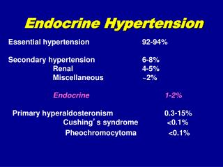
PHEOCHROMOCYTOMA • Arises from chromaffin cells in the sympathetic nervous system that release A, NA, and in some cases D • 0.1% of patients with diastolic hypertension have pheochromocytomas • In 50% of patients symptoms of are episodic/paroxysmal Symptoms during or following paroxysms: headache, sweating, facial pallor, cold and moist hands, forceful heartbeat with or without tachycardia, anxiety or fear of impending death, tremor, seizures, fatigue or exhaustion, nausea and vomiting, abdominal or chest pain, visual disturbances • Symptoms between paroxysms: increased sweating, heat intolerance, cold hands and feet, weight loss, constipation, wide fluctuations of blood pressure, postural hypotension
With time attacks usually increase in frequency but do not change much in character. • Glycosuria after an attack! ( glycogenolysis, insulin release). • Paroxysms may be induced by deep palpation of the abdomen • Typically, commonly used antihypertensive drugs are ineffective Location • Over 95% of pheochromocytomasare found in the abdomen, and 85% of these are in the adrenal. • Chest: heart, posterior mediastinum • Multiple tumours in less than 10% of adults • Tumours are usually small (< 100 g) • Incidence of malignant tumours: 10% • Complications of hypertension are common: hypertensive retinopathy or nephropathy, congestive heart failure, CVA, MI. • Common causes of death: MI, CVA, arrhythmias, irreversible shock, renal failure, dissecting aortic aneurysm.
Hormonal assessment • Plasma catecholamines • Urinary catecholamines • Urinary metoxycatecholamines • Urinary VMA (vanillylmandelic acid) Glucagon test: 1 mg i.v., phentolamine (Regitine) should be available to terminate the induced episode. Sensitivity: 90%. Clonidine suppression test: 0.3 mg of clonidine p.o. 2-3 hrs. before sampling of blood for plasma NA level: no reduction of plasma NA Trial of phenoxybenzamine (Dibenzyline): 2-receptor blocker Localization of tumour • CT or MRI imaging (bright image with T2-weighting) • Scintigraphy: 131I-metaiodobenzylguanidine (MIBG) • Venous catheterization for catecholamines assessment
Management Medical preoperative preparation: • Phenoxybenzamine (Dibenzyline) propranolol when marked tachycardia or arrhythmias • Prazosin propranolol • Labetalol Treatment of attacks: • Phentolamine (Regitine) 5-10 mg i.v. • Sodium nitroprusside – i.v. infusion Surgery: • Caution: induction of anesthesia • Phentolamine or sodium nitroprusside i.v. infusion • After tumour removal: blood volume expansion with whole blood, plasma, or other fluids
RENOVASCULAR HYPERTENSION • The most common cause of renin-dependent hypertension • The most common correctable cause of secondary hypertension (present in 1-4% of patients with hypertension) • Causes: • Atherosclerosis, • Fibromuscular hyperplasia, • Parenchymal lesions, hydronephrosis
When renovascular hypertension should be suspected? • Severe hypertension (diastolic pressure > 120 mmHg with either progressive renal insufficiency or refractoriness to agressive medical therapy (particularly in a smoker or with other evidence of occlusive arterial disease); • Accelerated or malignant hypertension with grade III or grade IV retinopathy; • Moderate to severe hypertension in a patient with diffuse atherosclerosis or a detected assymetry of kidney size; • An acute elevation in plasma creatinine level in a hypertensive patient that is either unexplained or follows therapy with an ACE inhibitor;
An acute rise in blood pressure over a previously stable baseline; • A systolic-diastolic abdominal bruit; • Onset of hypertension below age 20 or above age 50; • Moderate to severe hypertension in patients with recurrent acute pulmonary oedema; • Hypokalemia with normal or elevated plasma renin levels in the absence of diuretic therapy; • A negative family history of hypertension.
Diagnosis • Renal arteriography – a „golden standard • DSA – digital subtractive angiography • Captopril stimulation with measurement of PRA: exagerrated induction of reactive hyperreninemia • Captoptil renoscintigraphy: 90% sensitivity and specificity • Doppler ultrasound • MRI imaging • Spiral CT scan Treatment • Anatomic correction: surgery, angioplasty (PTCA) • Selective venous sampling for PRA (ratio affected kidney : contralateral kidney > 1.5 indicates functional abnormality) before anatomic correction • Medical treatment: ACE inhibitors, AT1 receptor antagonists particularly effective; beta-blockers, Ca channel blockers, methyldopa.