The Human Heart
The Human Heart. Heart Facts!. Your body contains about 5-6 L of blood Heart beats about 100,000 times/day or 35 million times in a year !


The Human Heart
E N D
Presentation Transcript
Heart Facts! • Your body contains about 5-6 L of blood • Heart beats about 100,000 times/day or 35 million times in a year! • The aorta, the largest artery in the body, is almost the diameter of a garden hose. Capillaries, on the other hand, are so small that it takes ten of them to equal the thickness of a human hair. • Your heart is roughly the size of your fist.
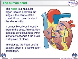
The basics… • muscular organ that pumps blood through vessels. • Pericardium: tough membrane that covers and protects the heart. • four chambers: • right/left atrium (contract at same time). • Right /left ventricles (contract at same time) • Blood enters atria and leaves through ventricles. • Double pump • Cardiac cycle (each heartbeat is called this)
Lungs Body cells Double-circulatory system… the right side of the system deals with deoxygenated blood. the left side of the system deals with oxygenated blood.
http://www.bhf.org.uk/swfs/hearthealth/structure_of_heart.swfhttp://www.bhf.org.uk/swfs/hearthealth/structure_of_heart.swf • left & right atria - upper chambers • thin walled; receive blood. • left & right ventricles - lower chambers • thick walled. • composed of cardiac muscle tissue • septumdivides two ventricles to prevent oxygen poor blood from right side of heart from mixing with oxygen rich blood of the left side.
Valves: • Control direction of blood flow inside the heart, allowing it to only flow one way. • atrioventricular valves (A-V valves)- between atrium and ventricles to prevent backward flow of blood; allow blood to flow from atria to the ventricles 1) right side: tricuspid; 3 flaps of tissue. 2) left side: bicuspid valve (mitral valve); 2 flaps of tissue. • semilunar valves - valves in heart at start of 2 major arteries leaving each ventricle. - allow blood to flow from ventricles to arteries. - open: blood flows into arteries. - closed: prevent blood from flowing back into ventricles. Right side: pulmonary valve Left side: aortic valve • “lub-dup” sound of heartbeat caused by opening/closing of these valves. (lub – A-V; dub-semilunar)
Heart Anatomy: • http://www.nucleusinc.com/animation2.php • Valve Functionhttp://www.medmovie.com/p_in_interactives.htm
Heartbeat Cycle: • The heart is considered a 2 phase pump because: 1) there is a period of relaxation and a period of contraction. 2) the atria contract first, then the ventricles. 3) the right side moves blood to the lungs, the left side moves blood to the rest of body. ** at rest, the heart pumps 60-80 times/minute!
Diastole (period of relaxation) • Blood flows from the veins into the heart chambers • Systole (period of contraction) • The atria briefly contract and fill the ventricles with blood. • Then the ventricles contract and propel blood out.
The two largest veins in your body are • 1. Superior vena cava: carries deoxygenated blood from the head /upper body back to the right atrium. • 2. Inferior vena cava: carries deoxygenated blood from the legs/lower body back to the right atrium.
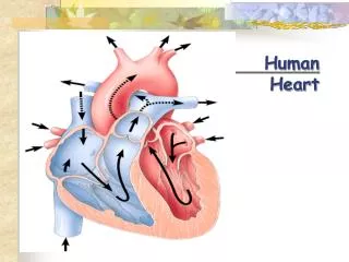
Blood Flow Through The Heart: • 1. Deoxygenated blood from the superior vena cava and inferior vena cava enter the right atrium. • 2. The right atrium releases blood through the tricuspid valve into the right ventricle. • 3. The right ventricle pushes the blood through the pulmonary valve into the pulmonary arteries. • 4. The pulmonary arteries take the blood to the left and right lungs.
5. In the lungs the blood flows through capillaries which allow for oxygen to enter the blood, and release carbon dioxide through the thin walled alveoli. 6. The now oxygen rich blood flows from each lung back to the heart via two sets of pulmonary veins. 7. These veins enter the left atrium. 8. The left atrium empties blood through the mitral valve to the left ventricle. 9. The left ventricle pushes the oxygen rich blood through the aortic valve to the aorta. 10. The aorta branches into many arteries which carry the blood to all parts of the body. http://www.medmovies.com/mmdatabase/mediaplayer.aspx?Message=VG9waWNpZD0xNDQ7Q2xpZW50SUQ9NDQ7VmVybmFjdWxhcklEPTE%3D-aYaBodAlDm0%3D
Passage of Blood Through the Heart • superior and inferior vena cava → right atrium → tricuspid valve → right ventricle → pulmonary semilunar valve → pulmonary trunk and arteries to the lungs → pulmonary veins leaving the lungs → left atrium → bicuspid valve → left ventricle → aortic semilunar valve → aorta → to the body. • *** FLOW CHART!!!!
Control of Heartbeat • Cardiac muscle has a built-in ability to contract. • 1) Sino-Atrial Node (S-A Node) - “pacemaker” • group of cells which control the beat of the heart by sending out electrical signals across the atria to cause them to contract. • 2) Atrio-Ventricular Node (A-V Node) • located between the right atrium and right ventricle • receives impulse from S-A node and sends electrical impulse across ventricles causing them to contract. • Electrocardiogram (ECG) – records electrical activity in the heart muscle; small spikes show contraction of atria while large spikes show contraction of ventricles. • http://health.howstuffworks.com/heart4.htm
Chemical Regulators • Eg. Page 318: You run to catch a bus • Faster cellular respiration= more CO2(blood) • Info goes to medulla oblongota (brain) • Release of noradrenaline=SA node fires rapid • When you sit down on the bus=medulla sends information through the nervous system • Medulla causes the release of acetylcholine which slows the firing of the SA node and therefore the heart rate slows.
Cardiac Output and Fitness • Cardiac output (CO) = the amount of blood pumped by the heart • Stroke volume( SV)=amount of blood forced out of the heart with each heartbeat (avg=70ml) • Heartrate (HR)=beats per minute (avg=70b/min) • CO=SV x HR • Eg. CO= 70 ml x 70 b/min • CO= 4900 ml/min
2 MAJOR DIVISIONS (Main pathways of circulation) • I Pulmonary Circulation • carries blood between heart and lungs. • function is to add oxygen to the blood and to remove carbon dioxide. • blood passes from right ventricle to lungs via pulmonary arteries and returns to left atrium via pulmonary veins. • Pulmonary arteries carry O2 poor blood. • Pulmonary veins carry O2 rich blood. • http://videos.howstuffworks.com/hsw/17110-the-circulatory-system-the-heart-video.htm
II Systemic Circulation • begins in the left ventricle of the heart. • blood is supplied to all other cells in the body with exception of the lungs. • supplies the body cells with oxygen and nutrients and takes away metabolic wastes and carbon dioxide. • http://www.youtube.com/watch?v=0jznS5psypI • http://www.wisc-online.com/Objects/ViewObject.aspx?ID=AP12704
Circulation of Lymph • Network of glands and vessels throughout your body. • Does not use a pump to circulate in the body. • Muscles in intestinal area and breathing muscles help fluid move. • Helps maintain the balance of fluids in the body. • Works with white blood cells to help guard against infection. • Blood fluid escapes through thin-walled capillaries into spaces between body tissues cells and forms part of interstitial fluid. • Lymph vessels pick up these fluids called lymph. • Lymph vessels join to form larger ducts that pass through lymph nodes. • Lymph nodes filter foreign substances such as bacteria and cancer cells. From the lymph before it is re-entered into the blood system. • Lymphocytes produced in lymph nodes – swell when sick.
Blood Pressure ** highest in the arteries and lowest in the veins! • the measurement of the pressure of blood created by the beating of the heart. • measures the pressure created by the expansion and retraction of arterial walls due to contraction and relaxation of heart. • measured using a sphygmomanometer. • normal blood pressure is 120/80. systolic diastolic
Systolic pressure: the highest pressure in the cardiac cycle, result of the ventricle contracting (heart at work). • Diastolic pressure: the lowest pressure in the cardiac cycle, just before the ventricle contracts (heart is at rest).
Hypertension: chronic high blood pressure caused by conditions that increase the volume of blood or reduce the elasticity of arteries. (stress, poor diet, lack of exercise, drugs, high cholesterol). • Atherosclerosis: “narrowing of the arteries” where a build up of cholesterol and fatty materials form plaque in the arteries. • Arteriosclerosis: natural part of the aging process in which arteries harden so that they are no longer flexible and will not expand.
Disorders of the Circulatory System • Anemia: lack of iron in the blood, low RBC count • Hemophilia: bleeders disease, lack of clotting factor • Heart attack: blood vessels around heart become blocked with plaque. Treatment includes: 1. clot busting drugs: must be administered immediately after stroke symptoms to work. 2. angioplasty: insertion of a tiny balloon to the clot site where it is inflated to force a vessel open. 3. coronary bypass surgery: use a healthy vessel from the patient’s body to create a new pathway around the blockage in a blood vessel near the heart. • Aneurysm: ballooning of a blood vessel, usually in the abdominal aorta or arteries leading to the brain; caused by high blood pressure and atherosclerosis) • Stroke - blood clot or ruptured artery interrupt blood flow to brain. • Hypertension • Arteriosclerosis
Preventing Heart Problems: • 1. Exercise – makes heart muscles stronger (heart beats more often when you exercise). • 2. Proper diet – overweight puts fat around heart; needs to work harder. • 3. Not smoking – nicotine causes blood vessels to narrow in size. • http://highered.mcgraw-hill.com/sites/0078617022/student_view0/brainpop_movies.html#