Human Heart
Human Heart. Made by: Marian Flaviu Stan Mariana Both on 8th grade. Human heart details.


Human Heart
E N D
Presentation Transcript
Human Heart Made by: Marian Flaviu Stan Mariana Both on 8th grade
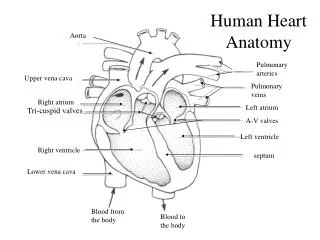
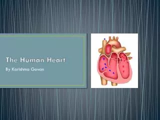
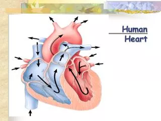
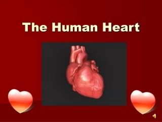
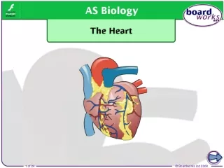
Human heart details • The human heart is a muscular organ that provides a continuous blood circulation through the cardiac cycle and is one of the most vital organs in the human body. The heart is divided into four main chambers: the two upper chambers are called the left and right Atrium and two lower chambers are called the right and left Ventricle.There is a thick wall of muscle separating the right side and the left side of the heart called the septum. Normally with each beat the right ventricle pumps the same amount of blood into the lungs that the left ventricle pumps out into the body. Physicians commonly refer to the right atrium and right ventricle together as the right heart and to the left atrium and ventricle as the left heart.
The electric energy that stimulates the heart occurs in the sinoatrial node, which produces a definite potential and then discharges, sending an impulse across the atria. In the atria the electrical signal moves from cell to cellwhile in the ventricles the signal is carried by specialized tissue called the Purkinje fiberswhichthen transmit the electric charge to the myocardium. • The human embryonic heart begins beating at around 21 days after conception, or five weeks after the last normal menstrual period (LMP). The first day of the LMP is normally used to date the start of the gestation (pregnancy). The human heart begins beating at a rate near the mother’s, about 75-80 beats per minute (BPM).
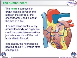
Structure • The human heart has a mass of between 250 and 350 grams and is about the size of a fist. • It is enclosed in a double-walled protective sac called the pericardium.Thedouble membrane of pericardium consist of the pericardial fluid which nourishes the heart and prevents shocks. The superficial part of this sac is called the fibrous pericardium. The fibrous pericardial sac is itself lined with the outer layer of the serous pericardium (known as the parietal pericardium). This composite (fibrous-parietal-pericardial) sac protects the heart, anchors its surrounding structures, and prevents overfilling of the heart with blood. The inner layer also provides a smooth lubricated sliding surface within which the heart organ can move in response to its own contractions and to movement of adjacent structures such as the diaphragm and lungs.
The outer wall of the human heart is composed of three layers. The outer layer is called the epicardium, or visceral pericardium since it is also the inner wall of the (serous) pericardium. The middle layer of the heart is called the myocardium and is composed of muscle which contracts. The inner layer is called the endocardium and is in contact with the blood that the heart pumps. Also, it merges with the inner lining (endothelium) of blood vessels and covers heart valves. • The human heart has four chambers, two superior atria and two inferior ventricles. The atria are the receiving chambers and the ventricles are the discharging chambers.
Functioning • Blood flows through the heart in one direction, from the atria to the ventricles, and out of the great arteries, or the aorta for example. Blood is prevented from flowing backwards by the tricuspid, bicuspid, aortic, and pulmonary valves. • The heart acts as a double pump. The function of the right side of the heart (see right heart) is to collect de-oxygenated blood, in the right atrium, from the body (via superior and inferior vena cavae) and pump it, via the right ventricle, into the lungs (pulmonary circulation) so that carbon dioxide can be dropped off and oxygen picked up (gas exchange). This happens through the passive process of diffusion.
On both sides, the lower ventricles are thicker and stronger than the upper atria. The muscle wall surrounding the left ventricle is thicker than the wall surrounding the right ventricle due to the higher force needed to pump the blood through the systemic circulation. Atria facilitate circulation primarily by allowing uninterrupted venous flow to the heart, preventing the inertia of interrupted venous flow that would otherwise occur at each ventricular systole. • Starting in the right atrium, the blood flows through the tricuspid valve to the right ventricle. Here, it is pumped out of the pulmonary semilunar valve and travels through the pulmonary artery to the lungs. From there, blood flows back through the pulmonary vein to the left atrium. It then travels through the mitral valve to the left ventricle, from where it is pumped through the aortic semilunar valve to the aorta and to the rest of the body. The (relatively) deoxygenated blood finally returns to the heart through the inferior vena cava and superior vena cava, and enters the right atrium where the process began.
Lifestyle and heart health • Obesity, high blood pressure, and high cholesterol can increase the risk of developing heart disease. However, half the number of heart attacks occur in people with normal cholesterol levels. Heart disease is a major cause of death. • It is generally accepted that factors such as exercise, diet, and overall well-being, including both emotional and physiological components, affect heart health in humans.