Download
1 / 49
500 likes | 629 Vues
Explore the vital role of the human heart in the circulatory system, including blood flow mechanisms and chamber functions. Delve into the structure, action, and control of the heart, encompassing cardiac cycle details and factors influencing heart rate and blood pressure regulation.

E N D
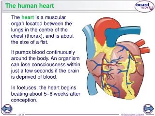
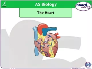
The HUMAN HEART The pump of the circulatory system
In the Human Circulatory System…… The flow of blood is maintained in 3 ways: 1 The pumping action of the heart arteries and capillaries 2 Contraction of skeletal muscles veins (aided by valves) 3 Inspiratory movements draws blood into the heart by the reduced pressure
How blood flows: the circulatory system Why is the blood blue on one side and red on the other?
How blood flows: the circulatory system Complete circulation consists of two pathways: pulmonary (lung) circulation & systemic (body) circulation
How blood flows: the circulatory system heart: coronary arteries & veins kidney: renal arteries & veins liver: hepatic artery & vein; gut/liver: hepatic portal vein (from gut to liver)
How blood flows: the circulatory system aorta: aortic arch & dorsal aorta venae cavae: superior (from head) & inferior (from body) Usually arteries are oxygenated, veins are deoxygenated
The mammalian heart lung heart liver stomach Our hearts are protected by our rib cages
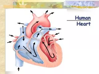
aorta superior vena cava pulmonary artery pulmonary veins left auricle coronary artery right auricle coronary vein right ventricle left ventricle inferior vena cava
Heart Structure & Action Structure of the Mammalian Heart - Pericardium: membrane around the heart - Cardiac muscles works (contracts) without rest - Coronary artery provides nutrients & oxygen to cardiac muscles Coronary veins carry away wastes and carbon dioxide from heart muscles - Septum divides the heart into right and left halves • The heart is divided into 4 chambers: • The left & right atria (auricles) and • The left & right ventricles
Atria • Left & right atria receive blood from veins and drain blood into the ventricles; • have thinner walls than ventricles • Right atrium receives deoxygenated blood from venaecavae (superior & inferior) - Left atrium receives oxygenated blood from pulmonary veins
Ventricles • pump blood to all parts of the body; have thicker,more muscular wallsthan the atria - Right ventricle pumps deoxygenated blood to the lungs through the pulmonary artery - Left ventricle pumps oxygenated blood into the aorta to deliver blood around the body (except the lungs) - Left ventricle has thicker wall than right ventricle because it has to pump blood all around the body, while the right ventricle only pumps blood to the lungs which are very close to the heart
Valves - enable blood to flow in only one direction: 1.Tricuspid valve: between right atrium and right ventricle 2.Bicuspid valve: between left atrium and left ventricle Both are attached by heart tendon to the muscular walls of ventricles in order to prevent the valves from being turned inside out 3.Semilunar valves: prevents blood flowing back from aorta and pulmonary artery tendon valve
Control of Heart Beat (Cardiac Cycle) All vertebrates are myogenic, i.e. the heart beat is initiated from within the heart muscle itself rather than an impulse from the brain or nervous system. The electronic heart pacemaker (SA node)
Nucleus Connective tissue Fibre with striations Cardiac muscle (LS)
Sino-atrial node (SA node) - located in wall of R A near the vena cava 'pace-maker' which determines the basic rate of heart beat: • wave of contraction from SA node both atria atrio-ventricular (AV) node Purkinje fibres (bundle of His) apex of ventricles contraction from ventricular apex upwards
Cardiac Cycle • Systole - heart contraction • Diastole - heart relaxation
Factors Modifying Heart Beat Cardiac output: Volume of blood pumped at heart beat x no. of beats/unit time - controlled by medulla oblongata by 2 centres: cardio-acceleratory centre (sympathetic) and cardio-inhibitory centre (parasympathetic)
During heavy exercise: CO2 pH chemoreceptor in carotid artery cardio-acceleratory centre heart beat (to remove more CO2) until CO2(pH) detected by carotid receptors cardio-inhitory centre heart beat
also: stretch receptors in aorta, carotid artery stimulated cardio-inhibitory centre heart beat stretch receptors in vena cava stimulated cardio-acceleratory centre heart beat
Maintenance & Control of Blood Pressure vasoconstriction: blood vessels narrowed blood pressure vasodilation: blood vessels dilated blood pressure Both controlled by vaso-motor centre in medulla oblongata arterioles in body (constrict/dilate) baroreceptors: pressure receptors in carotid artery detect blood pressure changes and relay impulses to the vaso-motor centre
examples: blood pressure baroreceptors vaso-motor centre sympathetic nerve arterioles vasoconstriction b p
blood pressure baroreceptors vaso-motor centre parasympathetic nerve arterioles vasodilation b p
Factors causing blood pressure increases: 1. CO2 blood pressure speed to deliver blood to heart remove more carbon dioxide 2. Hormones, e.g. adrenaline raises blood pressure
Pressure changes in the atria, ventricles & aorta during one cardiac cycle
Heart Disease - coronary heart disease 1. Coronary thrombosis - a blood clot blocking the coronary vessel Wall of artery Thrombus (clot) Plaque on inner wall of artery
Heart Disease - coronary heart disease 1. Coronary thrombosis - a blood clot blocking the coronary vessel 2. Atherosclerosis (hardening of the arteries) - barrowing of the arteries due to fat, fibrous, or salt deposits 3. Spasm - repeated contractions of the muscles in the coronary attery walls
Heart attack smooth lining blood flow restricted Cholesterol, Narrows the lumen in arteries, Decreases blood supply to organs artery blocked by blood clot fatty & fibrous deposits
The lymphatic system • It consists of lymph vessels with lymph.
Lymphatic System Tissue fluid and its formation • composition same as blood but without RBCs, platelets & proteins because they are too large to leak out of the capillaries - forms a link between blood and cells, providing a medium for exchange of materials between blood & cells
Lymph some tissue fluid returns to capillaries by osmosis while some (about 10%) goes into lymph capillaries; this fluid is now calledlymph Path: Blood lymph capillaries lymph vessels lymph ducts Blood - Lymph re-enters blood
At the arterial endof a capillary, liquid is forced out as tissue fluid which is similar to plasma in composition except its has no plasma proteins, platelets & RBCs. At the venous end, some fluid returns to blood while some enters lymph vessels which eventually join to a vein near the heart.
Lymph is driven by contraction of surroundingmuscles, aided by valves which enable one-way flow to the neck.
Lymph nodes - filter lymph passing through; with numerous WBCs to kill bacteria or neutralize toxins for bodily defence - during infection these nodes frequently swell - major sites of lymphocytes production
Movement of lymph through the lymphatic system: 1. Hydrostatic pressure 2. Muscle contraction 3. Inspiratory movement 4.Valves to ensure one-way traffic towards the heart
Functions of the lymphatic system 1 Transport of tissue fluid back into the blood circulation 2 Transport of fat from intestinal villi 3 As a bridge for the exchange of materials between capillaries and tissue cells 4 As a site for body defence (lymph nodes with WBCs)