Central Nervous System:
Central Nervous System:. Brain Spinal cord. The Brain. Performs the most complex neural functions Intelligence Consciousness Memory Sensory-motor integration Involved in innervation of the head. Organization of CNS.


Central Nervous System:
E N D
Presentation Transcript
Central Nervous System: • Brain • Spinal cord
The Brain • Performs the most complex neural functions • Intelligence • Consciousness • Memory • Sensory-motor integration • Involved in innervation of the head
Organization of CNS • Centrally located gray matter – neuron cell bodies, interneurons, unmyelinated fibers • Externally located white matter – myelinated fibers • Additional layer of gray matter external to white matter is the Cortex • Formed from neuronal cell bodies migrating externally • Located in cerebrum and cerebellum
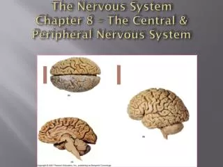
Basic Parts and Organization of the Brain • Divided into four regions • Cerebral hemispheres - Account for 83% of brain mass • Diencephalon – includes thalamus and hypothalamus • Brain stem - includes midbrain, pons, and medulla • Cerebellum – “little brain”
The Cerebral Hemispheres • Frontal section through forebrain • Cerebral cortex • Cerebral white matter • Deep gray matter of the cerebrum (basal ganglia) • Corpus Callosum – commissural fibers (white matter) which connects the two hemispheres
The Cerebral Hemispheres • Fissures – deep grooves, which separate major regions of the brain • Transverse fissure – separates cerebrum and cerebellum • Longitudinal fissure – separates cerebral hemispheres • Sulci - grooves on the surface of the cerebral hemispheres • Gyri - twisted ridges between sulci • Prominent gyri and sulci are similar in all people
Central sulcus Postcentral gyrus Precentral gyrus Parietal lobe Frontal lobe Parieto-occipital sulcus (on medial surface of hemisphere) Lobes, sulci, and fissures of the cerebral hemispheres. Lateral sulcus Occipital lobe Temporal lobe Transverse cerebral fissure Cerebellum Pons (a) Medulla oblongata Spinal cord Gyrus Cortex (gray matter) Sulcus White matter Fissure (a deep sulcus)
The Cerebral Hemispheres • Central sulcus separates frontal and parietal lobes • Bordered by two gyri • Precentral gyrus • Postcentral gyrus • Parieto-occipital sulcus - separates the occipital from the parietal lobe • Lateral sulcus - separates temporal lobe from parietal and frontal lobes • Deeper sulci divide cerebrum into lobes
The Cerebral Hemispheres • Lobes are named for the skull bones overlying them • Frontal • Parietal • Temporal • Occipital
The Cerebral Cortex • Home of our conscious mind • Composed of gray matter - neuronal cell bodies, dendrites, and short axons • Folds in cortex – triples its size • Approximately 40% of brain’s mass
The Cerebral Cortex - Functional Areas • Three general kinds of functional areas • Sensory areas • Association areas • Motor areas • Each of the major senses has a specific brain region called a primary sensory cortex • There are also multimodal association areas to process information
Motor areas Central sulcus Sensory areas and related association areas Primary motor area Premotor cortex Primary somatosensory cortex 3 1 2 Frontal eye field Somatic sensation 6 4 5 Somatosensory association area 7 Broca’s area (outlined by dashes) 8 Functional Areas Of The Cerebral Cortex Gustatory cortex (in insula) Taste Prefrontal cortex Wernicke's area (outlined by dashes) Working memory for spatial tasks 43 45 44 22 42 41 Executive area for task management 19 18 Primary visual cortex 22 17 47 Working memory for object-recall tasks 11 Vision Visual association area Solving complex, multitask problems Auditory association area Hearing Primary auditory cortex (a)
Primary Somatosensory Cortex • Located along the postcentral gyrus • Involved with conscious awareness of general somatic senses Motor areas Central sulcus Sensory areas and related association areas Primary motor area Premotor cortex Primary somatosensory cortex 3 1 2 Somatic sensation 4 5 Somatosensory association area 7 (a)
Primary Visual Cortex • On medial part of the occipital lobe • Largest of all sensory areas • Receives visual information that originates on the retina • First of a series of areas processing visual input 19 18 Primary visual cortex 17 Vision Visual association area (a)
Primary Auditory Cortex • Located at superior edge of the temporal lobe • Conscious awareness of sound • Impulses transmitted to primary auditory cortex Auditory association area Hearing Primary auditory cortex (a)
Olfactory Cortex • Olfactory nerves transmit impulses to the olfactory cortex • Provides conscious awareness of smells • Lies on the medial aspect of the temporal lobe 18 18 Visual association area 17 34 28 Olfactory bulb Primary visual cortex Temporal lobe Olfactory tract Fornix (b) Primary olfactory cortex
Gustatory Cortex • Involved in the conscious awareness of taste stimuli • Located on the “roof” of the lateral sulcus Gustatory cortex (in insula) Taste (a)
Motor areas Central sulcus Sensory areas and related association areas Primary motor area Premotor cortex Primary somatosensory cortex 3 1 2 Frontal eye field Somatic sensation 6 4 5 Somatosensory association area 7 Broca’s area (outlined by dashes) 8 Functional Areas Of The Cerebral Cortex Gustatory cortex (in insula) Taste Prefrontal cortex Wernicke's area (outlined by dashes) Working memory for spatial tasks 43 45 44 22 42 41 Executive area for task management 19 18 Primary visual cortex 22 17 47 Working memory for object-recall tasks 11 Vision Visual association area Solving complex, multitask problems Auditory association area Hearing Primary auditory cortex (a)
Motor Areas – Primary Motor Cortex • Controls motor functions • Located in precentral gyrus Motor areas Central sulcus Primary motor area Premotor cortex Primary somatosensory cortex 3 1 2 Frontal eye field 6 4 5 (a)
The Diencephalon • Forms the center core of the forebrain, primarily composed of gray matter • Surrounded by the cerebral hemispheres • Composed of three paired structures: thalamus, hypothalamus, epithalamus • Border the third ventricle
The Diencephalon • Forms the center core of the forebrain, primarily composed of gray matter • Surrounded by the cerebral hemispheres • Composed of three paired structures: thalamus, hypothalamus, epithalamus • Border the third ventricle Corpus callosum Septum pellucidum Fornix Choroid plexus Interthalamic adhesion (intermediate mass of thalamus) Thalamus (encloses third ventricle) Interventricular foramen Pineal body/gland (part of epithalamus) Corpora quadrigemina Hypothalamus Cerebral aqueduct Optic chiasma Midbrain Pituitary gland Arbor vitae Fourth ventricle Mammillary body Choroid plexus Pons Cerebellum Medulla oblongata Spinal cord
The Thalamus • Makes up 80% of the diencephalon • Contains approximately a dozen major nuclei • Act as relay stations for incoming sensory message • Every part of brain communicating with cerbral cortex relays signals through thalamic nuclei! • Is the “gateway” to the cerebral cortex
The Hypothalamus • Lies between the optic chiasm and the mammillary bodies • Pituitary gland projects inferiorly • Contains approximately a dozen nuclei • Main visceral control center of the body • The master gland’s master!!
The Diencephalon – The Hypothalamus • Functions include the following • Control of the ANS • Control of emotional responses • Regulation of body temperature • Regulation of hunger and thirst sensations • Control of behavior • Regulation of sleep-wake cycles • Control of the endocrine system • Formation of memory
The Diencephalon – The Epithalamus • Forms part of the “roof” (top) of the third ventricle • Consists of a tiny group of nuclei • Includes the pineal gland (pineal body) • Secretes the hormone melatonin • Under influence of the hypothalamus • Aids in control of circadian rhythm
The Brain Stem • Several general functions • Produces automatic behaviors necessary for survival • Passageway for all fiber tracts running between the cerebrum and spinal cord • Heavily involved with the innervation of the face and head • 10 of the 12 pairs of cranial nerves attach to it
The Brain Stem • Includes the midbrain, pons, and medulla oblongata
The Brain Stem – The Midbrain • The midbrain processes visual and auditory information and generates involuntary somatic motor responses • Has reticular activing system-arousal of the whole brain • Has nuclei for cranial nerves II and IV • Has ascending and descending tracts
The Brain Stem – The Midbrain • Lies between the diencephalon and the pons • Cerebral peduncles located on the ventral surface of the brain, contain pyramidal (corticospinal) tracts • Superior cerebellar peduncles - connect midbrain to the cerebellum
The Brain Stem – The Midbrain • Corpora quadrigemina (quad-ri-gemina) • The largest nuclei • Divided into the superior and inferior colliculi • Superior colliculi – nuclei that act in visual reflexes • Inferior colliculi – nuclei that act in reflexive response to sound
The Brain Stem – Dorsal View Figure 13.13c
The Brain Stem – The Pons • A “bridge” between the midbrain and medulla oblongata • Pons contains the nuclei of cranial nerves • V – Trigeminal nerve • VI – Abducens nerve • VII – Facial nerve • Motor tracts coming from the cerebral cortex • Pontine nuclei • Connect portions of the cerebral cortex and cerebellum • Send axons to cerebellum through the middle cerebellar peduncles
The Brain Stem – The Medulla Oblongata • The core of the medulla contains • Much of the reticular formation • Nuclei then influence autonomic functions • Cardiac center • Vasomotor center • The medullary respiratory center • Centers for hiccupping, sneezing, swallowing, and coughing
Functional Brain Systems – The Reticular Formation Figure 13.29
The Brain Stem – The Medulla Oblongata • Most caudal level of the brain stem • Is continuous with the spinal cord • Choroid plexus lies in the roof of the fourth ventricle • Cranial nerves VIII–XII attach to the medulla • External landmarks of medulla • Pyramids of the medulla lie on its ventral surface • Decussation of the pyramids - crossing over of motor tracts • Inferior cerebellar peduncles - fiber tracts connecting medulla and cerebellum
The Cerebellum • Located dorsal to the pons and medulla • Smoothes and coordinates body movements • Helps maintain equilibrium • Consists of two cerebellar hemispheres • Cortex – gray matter • Arbor vitae - internal white matter • Thick tracts connecting the cerebellum to the brain stem are superior, middle, inferior cerebellar peduncles
The Cerebellum • Composed of • Cortex – gray matter • Arbor vitae - internal white matter • Thick tracts connecting the cerebellum to the brain stem are • Superior cerebellar peduncles • Middle cerebellar peduncles • Inferior cerebellar peduncles • Fibers to and from the cerebellum are ipsilateral -run to and from the same side of the body
The Cerebellum • Cerebellum receives information from the cerebral cortex • On equilibrium • On current movements of • Limbs, neck, and trunk
Ventricles of the Brain • Expansions of the brain’s central cavity • Filled with cerebrospinal fluid • Lined with ependymal cells • Continuous with each other • Continuous with the central canal of the spinal cord
Ventricles of the Brain • Lateral ventricles – located in cerebral hemispheres • Horseshoe-shaped from bending of the cerebral hemispheres • Third ventricle – lies in diencephalon • Connected with lateral ventricles by interventricular foramen • Cerebral aqueduct – connects 3rd and 4th ventricles • Fourth ventricle – lies in hindbrain • Connects to the central canal of the spinal cord
Protection of the Brain • The brain is protected from injury by • The skull • Meninges • Cerebrospinal fluid • Blood-brain barrier
Cerebrospinal Fluid (CSF) • Formed in choroid plexuses in the brain ventricles • Choroid plexus is • Located in all four ventricles • Composed of ependymal cells and capillaries • Arises from blood - 500 ml/day
Cerebrospinal Fluid • Fills the hollow cavities of the brain and spinal cord • Provides a liquid cushion for the spinal cord and brain • Other functions • Nourishes brain and spinal cord • Removes wastes • Carries chemical signals between parts of the CNS
Cerebrospinal Fluid (CSF) Figure 13.32b
Blood Brain Barrier • Extensive impermeable capillaries & sinuses • Perivascular feet of astrocytes cover and wrap around capillaries and promote tight junction formation • Protects brain from hormones & circulating chemicals • Prevents most blood-borne toxins from entering the brain • Not an absolute barrier • Nutrients such as oxygen pass through • Allows alcohol, nicotine, and anesthetics through • Many glucose transporters Figure 9-6: The blood-brain barrier
Meninges • Functions of meninges • Cover and protect the CNS • Enclose and protect the vessels that supply the CNS • Contain the cerebrospinal fluid between pia and arachnoid maters
Meninges • Dura Mater • Strongest of the meninges • Composed of two layers: periosteal layer & meningeal layer • Arachnoid Mater • Located beneath the dura mater • Arachnoid villi - Project through the dura mater, allow CSF to pass into the dural blood sinuses • Pia Mater • Delicate connective tissue, clings tightly to the surface of the brain • Follows all convolutions of the cortex
The Spinal Cord • Functions of the spinal cord • Spinal nerves attach to it • Provides two-way conduction pathway • Major center for reflexes • Location of the spinal cord • Runs through the vertebral canal • Extends from the foramen magnum to the level of the vertebra L1 or L2
The Spinal Cord • Cervical and lumbar enlargements - where nerves for upper and lower limbs arise • Conus medullaris - the inferior end of the spinal cord • Cauda equina - collection of spinal nerve roots • Filum terminale - long filament of connective tissue, attaches to the coccyx inferiorly
Spinal Cord Segments • Indicate the region of the spinal cord from which spinal nerves emerge • Designated by the spinal nerve that issues from it • T1 is the region where the first thoracic nerve emerges