Download
1 / 26
260 likes | 278 Vues
Explore a comprehensive case study of B-cell lymphoma in a mule's third eyelids, from diagnosis to treatment outcomes and histopathology results. This detailed report covers clinical findings, diagnostic plans, differential diagnoses, immunohistochemistry results, and more.

E N D
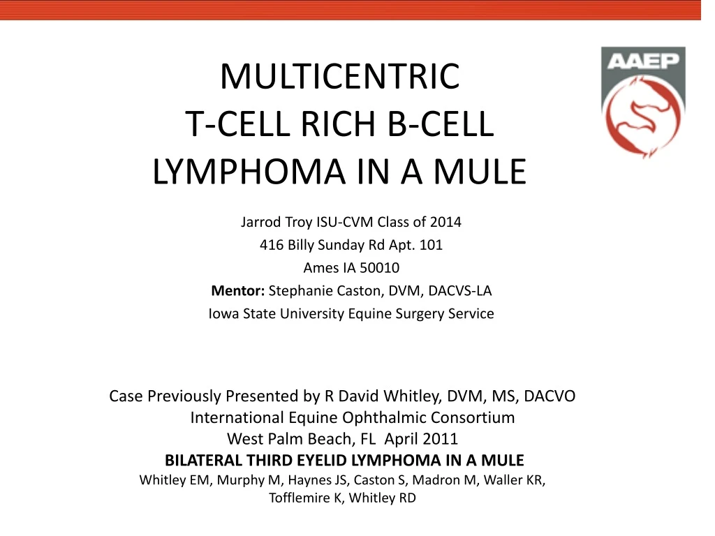
MULTICENTRICT-CELL RICH B-CELL LYMPHOMA IN A MULE Jarrod Troy ISU-CVM Class of 2014 416 Billy Sunday Rd Apt. 101 Ames IA 50010 Mentor: Stephanie Caston, DVM, DACVS-LA Iowa State University Equine Surgery Service Case Previously Presented by R David Whitley, DVM, MS, DACVO International Equine Ophthalmic Consortium West Palm Beach, FL April 2011 BILATERAL THIRD EYELID LYMPHOMA IN A MULE Whitley EM, Murphy M, Haynes JS, Caston S, Madron M, Waller KR, Tofflemire K, Whitley RD
SIGNALMENT • “Hank” • 25 year old castrated Mule • 495-kg (1090-lb) • Presented at Iowa State University Equine Surgery Service for bilateral surgical removal of third eyelids
HISTORY • 12/14/10: 6 week duration of progressive bilateral third eyelid swelling.
INITIAL CLINICAL FINDINGS • Bilateral bulbar and palpebral conjunctival thickening • Third eyelid protrusion • Mild bilateral exophthalmos • Patent nasolacrimal ducts • Mild ocular discharge
DIAGNOSTIC PLAN • Skull Radiographs • Dorsoventral & Lateral Oblique Views Collimated to Mid-Skull; Oblique Views Collimated to Orbits • Findings • Bilateral exophthalmos • Heterogeneous soft tissue masses (~7cm) rostroventral to globes • Guttural pouches partially air filled
PROBLEM LIST • Bilateral Bulbar and Palpebral Conjunctival Thickening • Third Eyelid Protrusion • Bilateral Exophthalmos • Mild Ocular Discharge • Guttural pouches partially air filled • Temperature Decreased • Tachypnea
DIFFERENTIAL DIAGNOSIS • Bilateral Bulbar/Palpebral Conjunctival Thickening • Foreign Body • Neoplasia • Blepharitis • Exophthalmos • Third Eyelid Protrusion • Trauma • Neoplasia • Blepharitis • Guttural Pouch Empyema • Bilateral Exophthalmos • Neoplasia • Orbital Cellulitis • Trauma • Mild Ocular Discharge • Neoplasia • Conjunctivitis • Exophthalmos • Trauma
DIFFERENTIAL DIAGNOSIS • Guttural pouches partially air filled • Trauma • Neoplasia • Guttural Pouch Empyema • Slightly Decreased Temperature • Cold Stress • Poor Perfusion • Trauma • Tachypnea • Stress • Pain • Pneumonia • Neoplasia
DIAGNOSIS • Presumptive Diagnosis • Severe, Bilateral Inflammation of Third Eyelid and Palpebral Conjunctiva • Possible Mass in the Guttural Pouch Area
TREATMENT PLAN • Palliative Therapy • Bilateral Surgical Removal of Third Eyelids • Eyelids were submitted for Histopathology • Home Treatment Instructions • Analgesia/ Anti-inflammatory • Phenylbutazone (4.4mg/kg, PO, SID for 7 days, then 1-2 gram as needed to decrease swelling or discomfort) • Topical antibiotic ointment • NEOMYCIN/POLYMYXIN B/BACITRACIN ZINC EYE OINT 3.5 (BID until tube is empty) • Clean discharge/blood from eyes with wet paper towel • Bloody discharge normal for 1-2 days post-op
OUTCOME 1 • “Hank” was discharged from hospital with Home Treatment Instructions • Third Eyelids were submitted for Histopathology
HISTOPATHOLOGY RESULTS • Both third eyelids, lacrimal gland, and adjacent conjunctiva were effaced by an infiltrative, non-encapsulated, poorly demarcated neoplasm • The neoplasm was composed of densely packed with a pleomorphic population of round cell sheets. • Mitotic figures are 3-5 per 400X field. • Moderate Anisocytosis/Anisokaryosis.
HISTOPATHOLOGY RESULTS • Neoplastic cells do not extend into the overlying, intact conjunctival epithelium. • Neoplastic cells extend to many tissue margins. • Microscopic Diagnosis • Third Eyelid Conjunctival Lymphoma • Immunohistochemical staining was requested to identify cell lineage
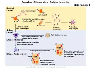
IMMUNOHISTOCHEMISTRY RESULTS • Immunohistochemical Staining • CD79a Positive Cells Indicate B-Lymphocyte Lineage • CD-3 Positive Cells Indicate T-Lymphocyte Lineage • Results: • Predominant population of cells were B-lymphocytes (CD79a-positive) • Small number of scattered T-lymphocytes (CD-3-positive) • Diagnosis • B-cell lymphoma CD79a-Positive Cells(B-Cells)
OUTCOME 2 • 12/29/12: “Hank” was readmitted to ISU Equine Surgery Service • Decreased condition • Weight Loss • Swelling at site of third eyelid removal • History • 12/14/12: Bilateral Third Eyelid Removal • Biopsy/Histopathology of Third Eyelids • Dx: B-Cell Lymphoma
CLINICAL FINDINGS • Physical Examination • Bilateral lower eyelid/conjunctival swelling. • Bilateral ocular discharge • Poor dilation and inability to examine fundus of the Left Eye • Solid vitreous face and posterior lens luxation of Right Eye • Submandibular Lymph Nodes enlarged • Multiple movable, semi-firm masses at Thoracic Inlet/Pelvic Area - Not noted at previous exam • Mild inspiratory stridor at rest
DIAGNOSTIC PLAN • Rectal Exam • No Abnormalities noted • Abdominocentesis • Slightly cloudy • Protein = 2.2 g/dL • Neurology Exam • No Abnormalities noted • CBC • Slight Anisocytosis of RBC • No other abnormalities noted • Endoscopy of Upper Airway • Arytenoids obscured by ventral displacement of the roof of the pharynx • Purulent material found in the Left Guttural Pouch Lateral Compartment. • Unable to enter Right Guttural Pouch due to Swelling/Scarring Due to worsening of clinical signs the owner elected for humane euthanasia and necropsy
NECROPSY RESULTS-GROSS • Orbit Conjunctiva • Bilaterally Swollen approximately 3cm x 2cm • Right Guttural Pouch • 200-mL of thick, white/yellow exudate • Left Guttural Pouch • Moderate amount of friable material. • Multiple 1-10mm nodules caudal to pouch • Cranial Mediastinal Lymph Node • Enlarged and moderately firm. Diameter 10cm
NECROPSY RESULTS-GROSS • Left Caudal Lung Lobe • White 7cm nodule • On cut surface, there were areas of concentric thickening around luminal structures • Mesentery • Pedunculated 10-15cm diameter white/yellow mass • Multiple non-pedunculated, white/yellow masses were also present. • Gross Morphological Diagnoses • Multifocal Lymphadenopathy • Guttural Pouch Empyema
NECROPSY RESULTS HISTOPATHOLOGY • Pituitary Gland (Pars Distalis & Pars Intermedia) • Expanded by well-demarcated, non-encapsulated infiltrative neoplastic mass of densely packed round cell sheets • High number of mitotic figures (28/10hpf) and abnormal nuclei & nucleoli changes • Lymph Node • Diffusely enlarged, with increased numbers of germinal centers; large lymphocytes expanding germinal centers • Lung Parenchyma, Palpebral Conjunctiva, Eye, & Adipose Tissue • Tissues were expanded/effaced by Neoplastic Cells similar in morphology to those described in the Pituitary Gland
NECROPSY RESULTS IMMUNOHISTOCHEMISTRY • Pituitary Gland • Scattered cells stained positively for T-Lymphocytes (CD-3) and for B-Lymphocytes (CD79a). • Eyelid Mass • Approximately 90% of neoplastic cells were positive for B-Lymphocytes (CD79a) • A few scattered cells were positive for T-Lymphocytes (CD-3) CD-3 Positive (T-Lymphocyte) CD79a Positive (B-Lymphocyte)
FINAL DIAGNOSIS • T-Cell Rich B-Cell Lymphoma • Grave Prognosis • Most horses die or are humanely euthanized within 6 months of clinical onset • Common secondary tumor of palpebral conjunctiva • Often associated with multicentric lymphoma
T-CELL RICH B-CELL LYMPHOMA • Clinical Signs • Often Related to loss of organ function from Lymphocyte Infiltration/Mass Obstruction • Anorexia; Emaciation; Lymph Node Enlargement (~2/3 of Cases) • Ocular Manifestation of Multicentric Lymphoma • Infiltration of upper, lower and third eyelids, conjunctiva, orbit, and globe. Retinal Detachment. Common secondary tumor site for lymphoma
T-CELL RICH B-CELL LYMPHOMA • Treatment Options • Limited • Transient improvement in generalized forms • Cytotoxic drugs, immunomodulators, corticosteroids • Long term response is poor • Surgical resection of tumors • Sometimes curative or significantly prolong survival • Humane euthanasia
FURTHER READING • Germann SE, Richter M, Schwarzwald CC, Wimmershoff J, Spiess BM. Ocular and multicentric lymphoma in a young racehorse. Vet Ophthalmol. Sep 2008;11 Suppl1:51-56 • Meyer J, Delay J, Bienzle D. Clinical, laboratory, and histopathologic features of equine lymphoma. Vet Pathol. Nov 2006;43(6):914-924. • Reed S, Bayly W, Sellon D. Equine Internal Medicine. 2nd ed: Saunders; 2004. • Barnett K, Crispin S, Lavach J, Matthews A. Equine Ophthalmology.2nd ed: Saunders; 2004. • Kelley LC, Mahaffey EA(1998). Equine Malignant Lymphomas: Morphologic and Immunohistochemical Classification. Vet Pathol 35: 241
ACKNOWLEDGEMENTS • Mentor Stephanie Caston, DVM, DACVS-LA • Pictures Provided By Elizabeth Whitley, DVM, PhD, DACVP R. David Whitley, DVM, MS, DACVO Stephanie Caston, DVM, DACVS-LA Kenneth Waller, DVM • Pathology Elizabeth Whitely, DVM, PhD, DACVP Joseph S. Haynes, DVM, PhD, DACVP Molly D. Murphy, DVM, PhD • Radiology Kenneth Waller, DVM