Immune System and Endocrine Disorders
Immune System and Endocrine Disorders. Immunology Unit Department of Pathology King Saud University. Objectives. To understand that immune system can target either single or multiple organs To know that autoimmunity against endocrine glands can cause endocrine dysfunction

Immune System and Endocrine Disorders
E N D
Presentation Transcript
Immune System and Endocrine Disorders Immunology Unit Department of Pathology King Saud University
Objectives • To understand that immune system can target either single or multiple organs • To know that autoimmunity against endocrine glands can cause endocrine dysfunction • To understand various cellular and humoral auto-immune mechanisms involved in different endocrine disorders • To know about the endocrine disease associations
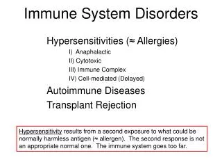
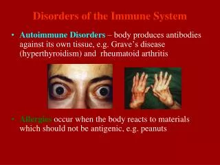
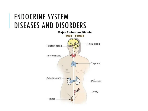
Immune response against endocrine glands Is organ-specific autoimmunity Both cellular and antibody mediated immune responses are involved Manifestations are usually related to the organ involved (Single or multiple organ involvement)
Autoimmune polyendocrine syndrome type • It is charaterized by multiple endocrine glands dysfunction as a result of autoimmunity and is associated with: • Hypoparathyroidism (Hypocaclaemia) • Addison’s disease (Hypoglycemia, hypotension) • Hypothyroidism • Hypogonadism and infertility • Vitiligo (depigmentation of the skin) • Alopecia(baldness) • Malabsorption • Pernicious anemia • Chronic active (autoimmune) hepatitis • Diabetes (Type 1)
Type I Diabetes: Pancreatic β cells express abnormally high levels of MHC I and MHC II (?) Normal PancreasPancreaswith Insulitis
Islet autoantigen Type 1 diabetes is T cell mediated • Infiltrating CD4+, CD8+ T cells • Anti-T cell therapies are effective • Islet cell autoantibodies disease CD4 T-cell CD8 T-cell TCR CD4 Treg Epitope HLA II HLA I APC Type 1 Diabetes: Cellular, Molecular & Clinical ImmunologyEdited by George S. EisenbarthOnline Edition Version 3.0
Type 1Diabetes mellitus(T1DM) • Predisposition • Genetic (HLA DRB, DQA, DQB) • Viral infections • Stress • Environmental exposure - exposure to certain chemicals or drugs • 10% chance of inheriting if first degree relative has diabetes • Most likely to be inherited from father
Type I Diabetes • Four auto-antibodies are markers of beta cell autoimmunity in type 1 diabetes: • Islet Cell Antibodies (ICA), against cytoplasmic proteins in the beta cell found in 75-90% patients • Antibodies to Glutamic Acid Decarboxylase 65 (GAD65) in 80% of patients • Insulin Auto-antibodies (IAA) is the first marker found in 70% of children at the time of diagnosis • IA-2A, (Insulinoma associated 2 auto-antibodies) to protein tyrosine phosphatasefound in 54-75% of patients
Prediction of TIDM
Limitations • Auto-antibodies may disappearmonths or years later without the development of diabetes • Since insulin-treated patients develop insulin antibodies, analysis of IAA is not useful in insulin-treated patients • Antibodies may be transferred trans-placentally to infants of type 1 diabetic mothers so caution must be used for interpretation
Anti-insulin antibodies • Anti-insulin antibodies either of IgG and/or IgM class against insulin are elevated and this may make insulin less effective or neutralize it IgG: is the most commontype of anti-insulin antibodyIgM: may cause insulin resistanceIgE: may be responsible for allergic reactions
Disease associations • About 10% patients with Type 1 diabetes are prone to other autoimmune disorders such as: • Addison’s disease • Pernicious anemia • Graves’ disease • Hashimoto’s thyroiditis
Thyroid autoimmunity • Hypothyroidism • Hashimoto’s disease • Atrophic thyroiditis • Hyperthyroidism • Graves’ disease
Chronic Lymphocytic Thyroiditis(Hashimoto’s Thyroiditis) • Male: Female ratio is 1:3 • Associated with HLA-B8 • Symptoms of hypothyroidism • Fatigue • Increased sensitivity to cold • Unexplained weight gain • Dry skin • Hoarseness • Puffy face • Thinning hair
Hashimoto’s Thyroiditis • Frequently affects middle-aged women • Individuals produce auto-antibodies and sensitized TH1 cells specific for thyroid antigens. • It is a delayed type of hypersensitivity response and is characterized by: • An intense infiltration of the thyroid gland by lymphocytes, macrophages, and plasma cells, which form lymphocytic follicles and germinal centers
Hashimoto’s-Pathogenesis • Sensitization of autoreactive CD4+ T-helper cells to thyroid antigens appears to be the initiating event. • The thyrocytesmay be destroyed by: • CD8+ cytotoxic T cell-mediated cell death:CD8+ cytotoxic T cells may cause thyrocyte destruction by either: • Perforin/granzyme granules • Engagement of death receptors (Fas) on the target cell • Cytokine-mediated cell death: CD4+ T cells produce inflammatory cytokines such as IFN-γ followed by recruitment and activation of macrophages and damage to follicles. • Binding of antithyroid antibodies: followed by antibody-dependent cell-mediated cytotoxicity (ADCC)
Hashimoto’s Thyroiditis • Lymphocytic infiltrate in the thyroid gland,with lymphoid follicle formation and fibrosis Normal thyroid histology Hashimoto’s throiditis with intense cellular infiltrates
Hashimotos’ thyroiditis • The inflammatory response causes: • A goiter, or visible enlargement of the thyroid gland • Antibodies are formed to a number of thyroid proteins, including: - Thyroglobulin - Thyroid peroxidase (both are involved in production of thyroid hormone)
Graves’ disease • Clinical Features: • Agitation • Sleep disturbance • Sweating • Palpitations • Muscle weakness • Weight loss despite increased appetite • Goiter • Tremor • Ophthalmopathy (Exopthalmos)
Graves’ Disease • Less common than • Hashimoto’s disease • Male: Female ratio 1:7 • Associated with HLA-B8 • Characterized by production of stimulating antibodies against TSH receptors
Graves' Disease • The production of antibodies in Graves' Disease is thought to arise by activation of CD4+ T-cells • Followed by B-cell recruitment into the thyroid • These B-cells produce antibodies specific to the thyroid antigens
Graves’ Disease • The thyrotropin receptor (Thyroid Stimulating Hormone (TSH) receptor) is the antigen for TSH receptor antibodies (TRAbs) • There are three types of TSH receptor antibodies: • Activating antibodies (associated with hyperthyroidism) • Blocking antibodies (associated with thyroiditis) • Neutral antibodies (no effect on receptor
Addison’s Disease • Female: Male ratio : 4:1 • Susceptibility genes: • HLA-DR3 and/or DR4
Primary adrenal insufficiency:Symptoms & Physical findings • Weakness • Weight loss • Poor appetite -Confusion • Hyper-pigmentation • Hypotension • Weak pulse • Shock
Autoimmune Adrenocortical Failure, or Addison's disease It develops as a consequence of autoimmune destruction of steroid-producing cells in the adrenal gland The major auto-antigen is 21-hydroxylase An enzyme involved in the biosynthesis of cortisol and aldosterone in the adrenal cortex
Autoimmune Adrenalitis • Humoral Immunity: • Autoimmune adrenalitis is characterized by the presence of serum antibodies against • 21-hydroxylase • 17-alpha-hydroxylase • The serum concentration of auto-antibodies, specifically against 21-hydroxylase correlates strongly with the degree of adrenal dysfunction • Adrenal insufficiency becomes clinically evident only after at least 90 percent of the cortex has been destroyed
Autoimmune Adrenalitis • Cellular Immunity: • The presence of lymphocytic infiltration in the adrenal glands • A Th1 response characterized by the production of Interferon-gamma • An antigen-specific T helper cell-driven process with cytotoxic T lymphocytes and activated macrophages mediate the destruction of the adrenal cortex
Gonads Autoimmune oophoritis (Inflammation of the ovaries) Autoimmune orchitis: Testicular pain involving swelling, inflammation and infection Pituitary gland Lymphocytic hypophysitis: Inflammation of the gland (autoimmune) resulting in decreased production of one or more hormones by the pituitary gland
Take home message • Either single or more than one endocrine glands may be targeted by immune system in a particular patient • Immunological damage to endocrine glands is mediated by autoimmune reactions • Endocrine dysfunction associated with immunological injury in majority of cases is due to end organ failure except in Graves’s disease. • Patients suffering from a particular immune mediated endocrine disorder frequently harbor antibodies against other endocrine glands.