Download
1 / 11
110 likes | 253 Vues
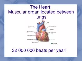
The Heart:. Muscular organ located between lungs. 32 000 000 beats per year!. structure. Composed of myocardium (cardiac muscle tissue) Inside the heart is lined with endocardium (squamous epithelial)

E N D
The Heart: Muscular organ located between lungs 32 000 000 beats per year!
structure • Composed of myocardium (cardiac muscle tissue) • Inside the heart is lined with endocardium (squamous epithelial) • Outside of heart is lined with pericardium which forms pericardial sac (for heart lubrication) • The heart is divided into two halves by the septum
Two halves of the heart are divided into 4 chambers • Blood flow through the heart is controlled by valves • The atrio-ventricular valves are held in place by chordae tendinae • The semi lunar valves direct flow out of the heart (resemble a half moon)
Function • The heart acts as a double pump • The right side of the heart pumps blood to the lungs (called the pulmonary circuit) • The left side pumps blood to the rest of the body (called systemic circuit) • This is why the left side of the heart is larger (has to pump blood farther)
Pathway of blood • Venae cavae (inferior and superior) to the right atrium • Right atrium to right ventricle • Right ventricle to pulmonary artery (to lungs/pulmonary veins and back to heart) • Left atrium to left ventricle • Left ventricle to aorta to the body
Systole – contraction: first atria contract, then ventricles • Diastole – relaxation • Sound: lub-dupp • Lub: atrioventricular valves close • Dupp: closing of semi-lunar valves • Heart murmur often due to ineffective valves (hear a “slush” sound instead of “lub”) this can cause back-flow in the heart
Blood pressure • Average = 120/80 mm Hg systole of left ventricle diastole of left ventricle • The ratio of the contraction and relaxations
Heart beat control • The heart beat is intrinsic, meaning it can beat independently of the central nervous system • The heart has its own pacemaker: the sino-atrial node • This is located in the upper dorsal wall of the right atrium
The SA node initiates the heart beat by causing the atria to contract • The electrical impulse then reaches the atrio-ventricular node located at the base of the right atrium • The signal is then transmitted through the ventricles by purkinje fibres • This then causes the ventricles to contract
The heart is controlled by the nervous system at the medulla oblongota (brain stem) • Heart beat is sped up or slowed down depending on what your body requires • Electrocardiogram (ECG) shows voltage changes across the heart during contraction