Bronchiectasis
Bronchiectasis. SS Visser, Pulmonology Internal Medicine UP. CONTENTS. Definition Aetiology Pathogenesis Clinical manifestations Diagnosis Treatment. BRONCHIECTASIS. Definition: Abnormal and permanent dilation of bronchi. Focal or diffuse distribution


Bronchiectasis
E N D
Presentation Transcript
Bronchiectasis SS Visser, Pulmonology Internal Medicine UP
CONTENTS • Definition • Aetiology • Pathogenesis • Clinical manifestations • Diagnosis • Treatment
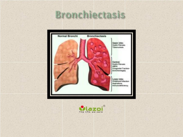
BRONCHIECTASIS Definition: Abnormal and permanent dilation of bronchi. Focal or diffuse distribution Clinical consequences – chronic and recurrent infection and Pooling of secretions in dilated airways. Classification: • Cylindrical (fusiform) • Saccular • Varicose
Measles and Pertussis Adeno & Influenza virus Bacterial infection with virulent organisms: S.aureus, Klebsiella Anaerobes Atypical mycobacteria Mycoplasma HIV Tuberculosis Fungi Aetiology: A. Infections-Micro-organisms
AETIOLOGY : IMPAIRED HOST DEFENCE • Local causes: Endobronchial obstruction • Generalised impairment: • 1. Immunoglobulin deficiency • 2. Primary ciliary disorders • 3. Cystic fibrosis
AETIOLOGY : NON-INFECTIOUS • Toxins or toxic substances NH3; gastric contents • Immune responses, ABPA • Inflammatory diseases: ulcerative colitis, rheumatoid arthritis, Sjögren syndrome. • -1-Antitrypsin deficiency • Yellow nail syndrome
CLINICAL MANIFESTATIONS • Persistent or recurrent cough with purulent sputum. • Haemoptysis • Initiating episode: Severe pneumonia, or insidious onset of symptoms or asymptomatic or non-productive cough – dry bronchiectasis in upper lobe, • Dyspnoea, wheezing – widespread bronchiectasis or underlying COPD. • Exacerbation of infection: Sputum volume increase, purulence or blood.
PHYSICAL EXAMINATION • Any combination of rhonchi, creps or wheezes. • Clubbing of digits. • Chronic hypoxaemia cor pulmonale R heart failure • Amiloidosis (rare)
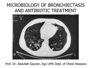
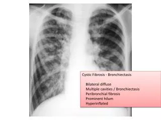
DIAGNOSIS - 1 • Clinical • Radiology: Chest XR: May be non-specific mild disease – normal XRC advanced disease – cysts + fluid levels peribronchial thickening, “tram tracks”, “ring shadows” CT Scan: Peribronchial thickening, dilated bronchioles. • Sputum culture: Pseudomonas aeuruginosa, H.influenzae.
DIAGNOSIS - 2 • Lung function: Airflow obstruction – FEV1 decreased. Air trapping - RV increased • Sweat test – increased sodium and chloride in cystic fibrosis • Bronchoscopy: Obstruction – foreign body, tumor. • Immunoglobulin • Cilia function and structure – Kartagener syndrome.
TREATMENT - 1 • 4 Goals: 1. Eliminate cause 2. Improve tracheo bronchial clearance 3. Control infection 4. Reverse airflow obstruction
TREATMENT - 2 • 1. Immunoglobulin 2. Antituberculous drugs 3. Corticosteroids (ABPA) 4. Remove aspirated material • Chest physical therapy • Mucolytics • Bronchodilators
TREATMENT - 3 • Antibiotics – short course, prolonged course, intermittent regular courses, inhalation. • Initial empiric Rx: Ampi, Amox, Cefaclor, Septran Ps.aeruginosa – Quinolone, aminoglycoside, 3rd generation cephalosporin, pipracillin. • Surgery: • Oxygen and diuretics • Lung transplant