Mol Genet 4
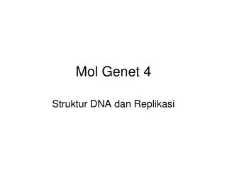
Mol Genet 4. Struktur DNA dan Replikasi. structure of DNA is a double-stranded, antiparallel helix. Antiparallel nature of the two DNA strands. ( B ) The double helical structure of DNA.

Mol Genet 4
E N D
Presentation Transcript
Mol Genet 4 Struktur DNA dan Replikasi
structure of DNA is a double-stranded, antiparallel helix. • Antiparallel nature of the two DNA strands. • (B) The double helical structure of DNA.
The two strands are antiparallel because they have opposite directions for linking of 3′ carbon atom to 5′ carbon atom. The structure shown is a double-stranded trinucleotide whose sequence can be represented as: 5′ pCpGpT-OH 3′ (DNA strand on left) / 5′ pApCpG-OH 3′ (DNA strand on right) (where p = phosphodiester bond and -OH = terminal OH group at 3′ end). This is normally abbreviated by deleting the ‘p' and ‘OH' symbols and giving the sequence on one strand only (e.g. the sequence could equally well be represented as 5′ CGT 3′ or 5′ ACG 3′).
Genetic information is encoded by the linear sequence of bases in the DNA strands (the primary structure). Consequently, two DNA strands of a DNA duplex are said to have complementary sequences (or to exhibit base complementarity) and the sequence of bases of one DNA strand can readily be inferred if the DNA sequence of its complementary strand is already known. It is usual, therefore, to describe a DNA sequence by writing the sequence of bases of one strand only, and in the 5′ → 3′ direction. This is the direction of synthesis of new DNA molecules during DNA replication, and also of the nascent RNA strand produced during transcription (see below). However, when describing the sequence of a DNA region encompassing two neighboring bases (really a dinucleotide) on one DNA strand, it is usual to insert a ‘p' to denote a connecting phosphodiester bond [e.g. CpG means that a cytidine is covalently linked to a neighboring guanosine on the same DNA strand, while a CG base pair means a cytosine on one DNA strand is hydrogen-bonded to a guanine on the complementary strand (Figure 1.6)].
Intermolecular hydrogen bonding also permits RNA-DNA duplexes and double-stranded RNA formation which are important requirements for gene expression (see Box 1.1). In addition, hydrogen bonding can occur between bases within a single DNA or RNA molecule. Sequences having closely positioned complementary inverted repeats are prone to forming hairpin structures or loops which are stabilized by hydrogen bonding between bases at the neck of the loop (Figure 1.7A). Such structural constraints, which are additional to those imposed by the primary structure, contribute to the secondary structure of the molecule. Certain RNA molecules, such as transfer RNA (tRNA), show particularly high degrees of secondary structure (Figure 1.7B).
Note that in the case of hydrogen bonding in RNA-RNA duplexes and also in intramolecular hydrogen bonding within an RNA molecule, GU base pairs are occasionally found (Figure 1.7B). This form of base-pairing is not particularly stable, but does not significantly disrupt the RNA-RNA helix.