Download
1 / 46
860 likes | 3k Vues
Ankle Fractures. OTA Fracture Course. Significance of Ankle Fractures. Most common weight bearing fracture we will treat Incidence is increasing Something we will all enc ounter. Lateral Ligaments. Medial Ligaments. Syndesmosis. Instability.

E N D
Ankle Fractures OTA Fracture Course
Significance of Ankle Fractures Most common weight bearing fracture we will treat Incidence is increasing Something we will all encounter
Instability • Inability to keep talus perfectly positioned under plafond • Usually lateral direction (external rotation) • Primary stabilizer is the deep deltoid JBJS 78, 1024
How much is too much • 1 mm translation decrease contact area by 42% JBJS 58,356
Indications for surgery • Instability of talus • Dynamic incongruity • Plafond step-off • Static iincongruity
X-ray • AP, Lateral and Mortise views of the ankle • AP and lateral of tibia • Consider stress views
Overlap Tilt Anteroposterior View
Mortise View Overlap Tilt Medial clear space
Posterior malleolus fracture Subluxation of the talus Distal fibula fx Lateral View
Classification • Lauge-Hansen • Danis-Weber (AO)
Danis-Weber Classification Doesn’t address the medial injury
Weber A • Supination-Adduction
Weber B • Supination-ER • With or without deltoid
Weber C • Pronation-ER • Pronation-Abduction
Medial Malleolar Fractures • Nondisplaced fractures may be treated nonoperatively • Displaced fractures • Isolated ?? ORIF • Part of bimalleolar pattern ORIF • Horizontal (tension) compression • Vertical (shear) antiglide plate
Lateral Malleolus Fractures Nonoperative management • 2-3 mm displacement • NO medial widening or syndesmotic injury • Cast or boot immobilization 6 wks • WBAT • Follow closely! • Stress view to ensure no medial injury ???
Surgical Indications • Bimalleolar / trimalleolar fractures • Bimalleolar equivalent • Syndesmotic disruption • Talarsubluxation
Implant ConsiderationsLateral Side • One-third tubular • “neutralization” plate laterally • “antiglide” plate posteriorly • 3.5 LCDCP on shaft
Posterior Malleolus • > 25% rule • Based on nothing • Fix if ANY posterior subluxation • A P or P A • Prone position very helpful
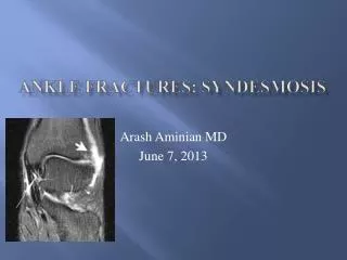
Biomechanics - Syndesmosis • Gardner 2006 • Compared fixation of syndesmosisvs posterior malleolus ORIF • Syndesmosis fixed= 40% of normal • Posterior malleolus fixed = 70% of normal
Maissoneuve Fracture • Fracture of proximal fibula • +/- medial malleolar fracture • Pronation-external rotation • Requires reduction and stabilization of syndesmosis
Maissoneuve Fracture • Fracture of proximal fibula • +/- medial malleolar fracture • Pronation-external rotation • Requires reduction and stabilization of syndesmosis
Syndesmosis Fixation • MUST test for Syndesmotic instability after fixation of lateral malleolus • Have bone hook on table to check stability, Cotton Test
Syndesmosis Controversies No significant difference !! 1 vs. 2 screws 3.5 vs. 4.5 screws 4 cortices vs. 3 Two hole plate Tightrope The key is the REDUCTION !!
Biomechanics • Complex motion • 5x body weight in stance • At least 10 degrees of dorsiflexion is needed for normal gait • 1 mm of lateral talar shift decreases tibiotalar surface contact up to 40%
MM LM Talus INVERSION Weber A Tibia IOM Fibula Tib-Fib Lig Delt Lig Talo-Fib Lig
MM LM Talus EVERSION Weber B Tibia IOM Fibula Tib-Fib Lig Delt Lig Talo-Fib Lig
MM LM Talus EVERSION Weber C Tibia IOM Fibula Tib-Fib Lig Delt Lig Talo-Fib Lig