Download
1 / 89
900 likes | 1.09k Vues
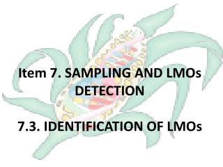
Item 7. SAMPLING AND LMOs DETECTION 7.3. IDENTIFICATION OF LMOs. Detection is required when :. By law in the country is required identification and/or labelling Mixtures between GMOs + non-GMOs Need to export to a country with strict legislation Need to verify non-GMOs shipments

E N D
Item 7. SAMPLING AND LMOs DETECTION7.3. IDENTIFICATION OF LMOs
Detection is required when: • By law in the country is required identification and/or labelling • Mixtures between GMOs + non-GMOs • Need to export to a country with strict legislation • Need to verify non-GMOs shipments • For environmental risk verifications
In the international market • Important focus differences between commercial blocks: • USA does not require identification and GM crops are easily set free into the environment • EU requires labelling with 0.9% treshold
“Historical” cans of GM tomato puree with long shelf life • First GM-food authorized 1994 • They were labelled… and people bought them!!! • The variety lost sensory characteristics and was retired from the market
Extraction of: Sampling Immunostrips ELISA plates Protein PCR end point DNA RTq-PCR
In order to detect proteins, specific antibodies are required. The antibodies are proteins with quaternary structure.
Lateral flow test, IMMUNOSTRIP Control line. Antibodies anti-IgG adsorbed Test line. Antibodies anti-antigen adsorbed Antibodies anti-antigen conjugated with enzyme
Lateral Flow test,IMMUNOSTRIP The antibodies are binded to their antigen in the sample and the complex antigen-antibody moves by capillarity towards the reaction lines.
Lateral Flow test,IMMUNOSTRIP Antigen binds to the antibodies that are in the test line. Free antibodies bind to the antibodies anti-Ig present in the control line.
Immunostrip procedure 1. Weigh 250 mg of fresh leaf (plantlet) 2. Insert the sample in the bag 3. Grind or crush the sample 5. Let stand for 10 minutes (vertical position) 4. Insert the immunostrip into the bag with SEB or MEB buffer. 6. Read results
Qualitative immunoassay in strip. Ej. Cry9C QuickStixTM Envirologix
Also by this technique can be detected CP4-EPSPS protein in samples with low % of transgenic maize
The first Ac is adsorbed to the plate ELISA: Enzyme-Linked-ImmunoSorbent Assay Format: DAS (Double Antibody Sandwich)
Sample that contains protein is added Protein ELISA: Enzyme-Linked-ImmunoSorbent Assay Formato: DAS (Double Antibody Sandwich)
Second antibody is added. It is conjugated with a enzyme Enzyme Antibody ELISA: Enzyme-Linked-ImmunoSorbent Assay Format: DAS (Double Antibody Sandwich)
Colorless compound Colorless compound Product Product Antibody Enzyme ELISA: Enzyme-Linked-ImmunoSorbent Assay Formato: DAS (Double Antibody Sandwich)
ELISA plate, second antibody congujated to alkaline phospatase
Quantitative Immunoassay (ELISA) Centrifugation to clarify samples Sample extraction. Preparation of ELISA plates
ELISA cont. Incubation at room temperature Sample application Substrate addition/ Colour development Spectrophotometric measurement - for quantification- Wash
1 2 3 1 2 3 A B C D E F G H A B C D E F G H Specificity assesment of antibodies in the ELISA test KIT CP4 EPSPS KIT Cry 1Ab/1Ac
1 2 3 4 5 A B C D E F G H Quantitative Analysis by ELISA in detection of protein CP4-EPSPS
Detection thresholds of three transgenic maize events CP4-EPSPS protein can be detected with high sensitivity in mixtures with low percentage of transgenic maize. Sensitivity for detection of CRY proteins is much lower.
Considerations:Immunochemical methods • Immunochemical methods can be realized in fast ways, in situ, or in laboratory, few equipment is required. • Strip based methods are only qualitative. • ELISA can be quantitative but: • Different levels of protein expression are reflected in different sensitivity levels • No reference materials recognized. • No agreement between quantification units (% in weight or protein concentration)
Sequence of interest Taq polymerase Synthesis of one copy PCR principles • Double chain opening (~90°C) –denaturation- • Primers that recognize specific sequences (50- 60°C) • Synthesis of template complementary chains (74°C) 3’ 5’ 5’ 3’ 3’ 5’ 5’ 3’
Amplification: Sequence of interest Taq Taq Taq Amplification Taq
Number of DNA molecules 1 2 4 8 16 32 64 128 256 512 1024 1,000,000 1,000,000,000 1,000,000,000,000 Number of PCR cycles 0 1 2 3 4 5 6 7 8 9 10 20 30 40
Theoretical PCR products Real Cycle number
Factors to consider • Specificity – primer design • Product length (DNA amplified fragment) • There are differences between qualitative and quantitative tests • Whether PCR is uniplex or multiplex • If the method is specific for a type of instrument
H G H P E T LOW 1. Exploration 2. Gene specific target Target specificity 3. Specific construct 4. Event specific target HIGH Primer design H Host genomic DNA P Promoter element (CaMV 35S) E Amplifying element G Gene of interest (Cry, EPSPS) T Terminator (NOS)
PCR for transgenic sequence detection Recombinant Gene 1 2 3 4 5 A B C D Amplicons
DNA of GM grains Genomic DNA Recombinant gene
Specific-specie primers recognize genomic DNA DNA of non-GM grains Genomic DNA Specific-GMO primers do not interact No amplification
Intrinsic factors of the sample that affect the amplification • Integrity of template DNA • Size of the amplicon • Presence of inhibitory substances • Humic substances • Proteins Others: • EDTA • NaOH • SDS and other detergents
1 Ladder 2A Seeds 2B Commercial nixtamal flour 3 Dough 4 Nixtamal flour 5 Tortilla 6 Tortilla chips 7 Corn chips 8 Dry Corn chips Load: 100ng Dye: SYBR Green
DNA Extraction • Extraction yield • Tissue • DNA purity • Quality for amplification
C C- HJ P M HM HMS HJS E S 225 pb A C- P HJ S HJS HM Es E M HMS 225 pb B HJS M HMS HM C- E HJ Es P S 225 pb Amplification – effect of the purity of the DNA template Amplification of a fragment of the Invertase gene.
MON810 B 10 1 C-4 C-3 0.1 C-2 M C-1 300 pb 100 pb Limit of Detection Detection of the specific event MON810 in different proportions (10%, 1% y 0.1%).C-1, negative control with BT11 maize seed DNA 100% transgenic; C-2, negative control with NK603 maize seed genomic DNA; C-3, negative control with chalqueño maize seed DNA; C-4, negative control without DNA. M, 50 bp ladder.
Identity verification: Restriction analysis Amplicons of CamV35S promoter and restriction products with Asp700. Lane 1: 50bp ladder, lanes 2 and 3: Bt176 control, lanes 4 and 5: canned corn grains, in 2% agarose gel.
PCR products 100 10 2 1 0.1 0.01 Cycle number Ct Ct = number of cycles needed for the amplification signal to be statistically different from the background signal m=-3.32 Log conc.
Number of DNA molecules 22 1 42 2 82 3 10 2 3.32 162 4 32... 64... 128... Number of PCR cycles 1 2 3 4 5 6 7
(-1/m) Efficiency = ([10 ]-1) 100 * Efficiency Results are accepted when the efficiency is higher than 95% (m = 3.45 a 3.3)
Some results in PCR real time Specificity of primers and probes designed for RTQ-PCR
Effect of the extraction system Figure 26. Standard curves of the events MON810 y Bt11 from DNA extracted with the commercial systems A, B and C. 1, curve of the event Bt11 with extraction system A. 2, curve of the event MON810 with extraction system A. 3, curve of the event MON810 with extraction system B. 4, curve of the event Bt11 with extraction system B. 5, curve of the event Bt11 with extraction system C. 6, curve of the event MON810 with extraction system C.