Antiarrhythmic Drugs
510 likes | 713 Vues
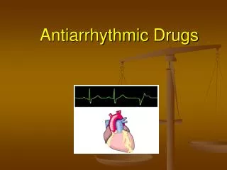
Antiarrhythmic Drugs. Arrhythmia. Heart condition where disturbances in Pacemaker impulse formation Contraction impulse conduction Combination of the two Results in rate and/or timing of contraction of heart muscle that is insufficient to maintain normal cardiac output (CO)

Antiarrhythmic Drugs
E N D
Presentation Transcript
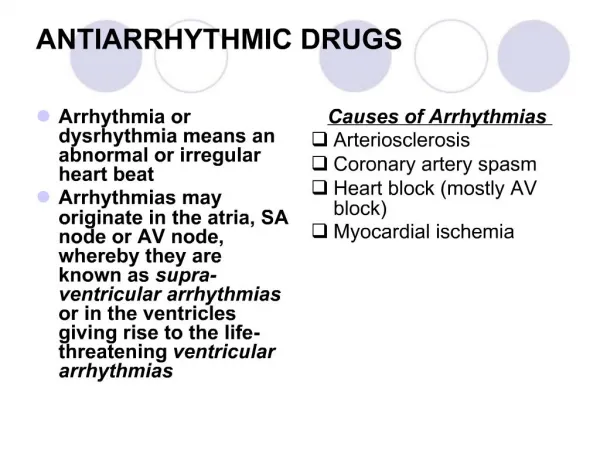
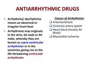
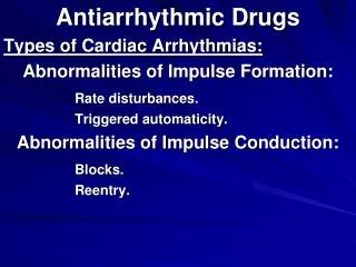
Arrhythmia • Heart condition where disturbances in • Pacemaker impulse formation • Contraction impulse conduction • Combination of the two Results in rate and/or timing of contraction of heart muscle that is insufficient to maintain normal cardiac output (CO) To understand how antiarrhythmic drugs work, need to understand electrophysiology of normal contraction of heart
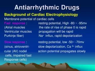
Action potential • Cardiac muscle cells are electrically excitable. • They show a spontaneous, intrinsic rhythm generated by specialized pacemaker cells located in the sinoatrial and atrioventricular nodes. • The cardiac cells also have an unusually long action potential, which canbe divided into five phases
Contraction of ventricles ECG (EKG) showing wave segments Repolarization of ventricles Contraction of atria
Differences between nonpacemaker and pacemaker cell action potentials • PCs - Slow, continuous depolarization during rest • Continuously moves potential towards threshold for a new action potential (called a phase 4 depolarization)
Normal heartbeat and atrial arrhythmia Normal rhythm Atrial arrhythmia AV septum
Causes of arrhythmias 1- Abnormal automaticity: • The SA node normally sets the pace of contraction for the myocardium, and other pacemakers are depolarized by impulses coming from the SA node. • if cardiac sites other than the SA node show enhanced automaticity, they may generate competing stimuli, and arrhythmias may arise.
2- Abnormalities in impulse conduction Impulses from higher pacemaker centers are normally conducted down pathways that bifurcate to activate the entire ventricular surface A phenomenon called reentry • if we consider a single Purkinje fiber with two conduction pathways to ventricular muscle. An impulse normally travels down both limbs of the conduction path. • However, if myocardial injury results in a unidirectional block, the impulse may only be conducted down Pathway 1 • This results in reexcitation of the ventricular muscle, causing premature contraction or sustained ventricular arrhythmia
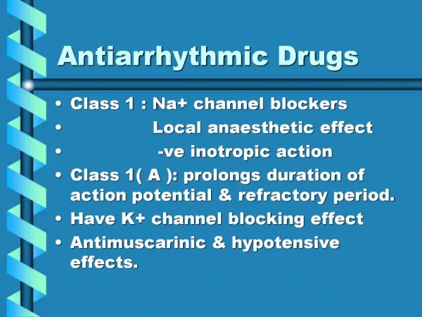
III. Class I Antiarrhythmic Drugs • Class I antiarrhythmic drugs act by blocking voltage-sensitive sodium channels via the same mechanism as local anesthetics. • The decreased rate of entry of sodium slows the rate of rise of Phase 0 of the action potential. • Class I antiarrhythmic drugs, therefore, generally cause a decrease in excitability and conduction velocity. A. Use-dependence • Class I drugs bind more rapidly to open or inactivated sodium channels than to channels that are fully repolarized following recovery from the previous depolarization cycle.
Class I drugs # Class IA agents • slow the rate of rise of the action potential (thus slowing conduction) • prolong the action potential, and increase the ventricular effective refractory period. • They have an intermediate speed of association with activated/inactivated sodium-channels and an intermediate rate of dissociation from resting channels. # Class IB drugs - They rapidly interact with sodium channels. # Class IC agents • markedly depress the rate of rise of the membrane action potential. • They bind slowly to sodium channels.
Quinidine • It is IA drug with Class III activity • Mechanism of action: • Quinidinebinds to open and inactivated sodium channels and prevents sodium influx, • thus slowing the rapid upstroke during Phase 0 . • It also decreases the slope of Phase 4 spontaneous depolarization and inhibits potassium channels. • Therapeutic uses: • used in AV-junctional, and ventricular tachyarrhythmias. Quinidineis used to maintain sinus rhythm after direct-current cardioversion of atrial flutter or fibrillation and to prevent frequent ventricular tachycardia. • Adverse effects: • Torsde de point, Asystole, Anticholinergic, cinchonism
Procainamide • Actions: This Class IA drug, a derivative of the local anesthetic procaine, shows actions similar to those of quinidine. • Pharmacokinetics: • It is acetylated in the liver to N-acetylprocainamide (NAPA). • NAPA has properties of a Class III drug. NAPA is eliminated via the kidney, and dosages of procainamidemay need to be adjusted in patients with renal failure. • Adverse effects: • reversible lupus erythematosus“like syndrome asystole or induction of ventricular arrhythmias. depression, hallucination, and psychosis.
Disopyramide • Actions: This Class IA drug shows actions similar to those of quinidine. • Disopyramidecauses peripheral vasoconstriction. • Used in the treatment of ventricular arrhythmias as an alternative to procainamideor quinidine. • Adverse effects: Disopyramideshows effects of anticholinergic activity (for example, dry mouth, urinary retention, blurred vision, and constipation).
Lidocaine • It is a Class IB drug. • Therapeutic uses: • Lidocaineis useful in treating ventricular arrhythmias arising during myocardial ischemia, such as that experienced during a myocardial infarction. • Pharmacokinetics: Lidocaineis given intravenously because of extensive first-pass transformation by the liver, • Adverse effects: Lidocainehas a fairly wide toxic-to-therapeutic ratio. It shows little impairment of left ventricular function and has no negative inotropic effect • CNS effects include drowsiness, slurred speech, paresthesia, agitation, confusion, and convulsions. • Cardiac arrhythmias may also occur.
Mexiletine and tocainide • These Class IB drugs have actions similar to those of lidocaine, and they can be administered orally. • Mexiletine is used for chronic treatment of ventricular arrhythmias associated with previous myocardial infarction. • Tocainideis used for treatment of ventricular tachyarrhythmias • Tocainide causes pulmonary fibrosis. FlecainideIt is a Class IC drug. • Therapeutic uses: • in treating refractory ventricular arrhythmias. It is particularly useful in suppressing premature ventricular contraction. • Flecainidehas a negative inotropic effect and can aggravate congestive heart failure. • Propafenone: Class IC similar flecainide.
Class II Antiarrhythmic Drugs (Beta-blockers) ►These drugs diminish Phase 4 depolarization, thus depressing automaticity, prolonging AV conduction, and decreasing heart rate and contractility. ► uses ♥ tachyarrhythmias caused by increased sympathetic activity. ♥atrial flutter and fibrillation ♥ AV-nodal reentrant tachycardia. ♥ Propranolol reduces the incidence of sudden arrhythmic death after myocardial infarction Metoprolol • it reduces the risk of bronchospasm. Esmolol • It is a very short-acting beta-blocker used for intravenous administration in acute arrhythmias that occur during surgery or emergency situations.
Class II Antiarrhythmic Drugs (Beta-blockers) • These drugs diminish Phase 4 depolarization, thus depressing automaticity, prolonging AV conduction, and decreasing heart rate and contractility. • Class II agents are useful in treating tachyarrhythmias caused by increased sympathetic activity. They are also used for atrial flutter and fibrillation and for AV-nodal reentrant tachycardia. [ • Propranolol reduces the incidence of sudden arrhythmic death after myocardial infarction • B. Metoprolol • it reduces the risk of bronchospasm. • C. Esmolol • It is a very short-acting beta-blocker used for intravenous administration in acute arrhythmias that • occur during surgery or emergency situations.
Class III Antiarrhythmic Drugs Amiodarone ☺ it contains iodine and is related structurally to thyroxine. ☺ Class I, II, III, and IV actions. Its dominant effect is prolongation of the action potential duration and the refractory period. ☺ Therapeutic uses: ♦ treatment of severe refractory supraventricular and ventricular tachyarrhythmias. ♦ Despite its side-effect profile, amiodaroneis the most commonly employed antiarrhythmic. ☺ Pharmacokinetics: • It has a prolonged half-life of several weeks, and it distributes extensively in adipose tissue. Full clinical effects achieved until 6 weeks
☺ Adverse effects: ► interstitial pulmonary fibrosis ► gastrointestinal tract intolerance ► tremor, ataxia, dizziness ► hyper- or hypothyroidism ► Liver toxicity ► photosensitivity ► neuropathy ► muscle weakness ► blue skin discoloration caused by iodine accumulation in the skin.
Sotalol ► class III antiarrhythmic agent and has potent nonselective beta-blocker activity. ► It prolongs both repolarization and duration of the action potential, thus lengthening the effective refractory period. Therapeutic uses: ► antiarrhythmic ► Beta-Blockers are used for long-term therapy to decrease the rate of sudden death following an acute myocardial infarction.
Class IV Antiarrhythmic Drugs • Class IV drugs are calcium-channel blockers resulting in a decreased rate of Phase 4 spontaneous depolarization. They also slow conduction in tissues that are dependent on calcium currents, such as the AV node . Verapamil and diltiazem • Verapamil shows greater action on the heart than on vascular smooth muscle • nifedipine, a calcium-channel blocker used to treat hypertension exerts a stronger effect on the vascular smooth muscle than on the heart. • Diltiazem is intermediate in its actions.
Actions: ►Verapamiland diltiazembind only to open, depolarized channels so they are therefore use-dependent ►verapamiland diltiazemslow conduction and prolong the effective refractory period in tissues that are dependent on calcium currents, as the AV node. ► They treat arrhythmias that must traverse calcium-dependent cardiac tissues.
☺ Therapeutic uses: ►Verapamiland diltiazemare more effective against atrial than ventricular arrhythmias. ► They are useful in treating reentrant supraventricular tachycardia and in reducing the ventricular rate in atrial flutter and fibrillation. ► They used to treat hypertension and angina. ☺ Adverse effects: ►Verapamiland diltiazemhave negative inotropic and contraindicated in patients with preexisting depressed cardiac function. Both ► drugs can also produce a decrease in blood pressure because of peripheral vasodilation an effect that is actually beneficial in treating hypertension.
►Digoxin • It decreases conduction velocity in the AV node. • Digoxinis used to control the ventricular response rate in atrial fibrillation and flutter. • At toxic concentrations, digoxincauses ventricular tachycardia and fibrillation which treated with lidocaineor phenytoin. ► Adenosine Mechanism of action inhibition of the slow inward calcium current and activates adenylatecyclase through A2 receptors in smooth muscle at high doses, the drug decreases conduction velocity, prolongs the refractory period, and decreases automaticity in the AV node. • Intravenous adenosine is the drug of choice for abolishing acute supraventricular tachycardia. It has low toxicity but causes flushing, chest pain, and hypotension. • short duration of action (15 sec).
Pacemakers Surgical implantation of electrical leads attached to a pulse generator 1- Leads are inserted via subclavicle vein and advanced to the chambers on the vena cava (right) side of the heart Two leads used, one for right atrium, other for right ventricle • Pulse generator containing microcircuit and battery are attached to leads and placed into a “pocket” under the skin near the clavicle • Pulse generator sends signal down leads in programmed sequence to contract atria, then ventricles • Pulse generator can sense electrical activity generated by the heart and only deliver electrical impulses when needed.
Heart Failure • the heart is unable to pump sufficient blood to meet needs of the body. • Underlying causes of HF include • arteriosclerotic heart disease • myocardial infarction • hypertensive heart disease • valvularheart disease • dilated cardiomyopathy • congenital heart disease.
Compensatory physiological responses in HF • 1- Increased sympathetic activity: decrease in B.P, activates the sympathetic which stimulates β1- receptors, increase HR & FC (+veino). vasoconstriction (α1-mediated) enhances venous return and increases cardiac preload. decline in cardiac function. • 2- Activation of the renin-angiotensinsystem • A fall in B.P releases renin, leaing to release of angiotensin II and aldosterone. • increased peripheral resistance and retention of sodium and water. Blood volume increases, and more blood is returned to the heart. • increase heart work and decline cardiac function.
3- Myocardial hypertrophy • The heart increases in size, and the chambers dilate and become more globula • Initially, stretching of the heart muscle leads to a stronger contraction of the heart. • However, excessive elongation of the fibers results in weaker contractions. This type of failure is termed systolic failure and is the result of a ventricle being unable to pump effectively. • If the adaptive mechanisms fail to maintain cardiac output, the HF is termed decompensated.
Treatment of CHF • 1- Inhibitors of the Renin-Angiotensin System • A. Angiotensin-converting enzyme inhibitors • ACE inhibitors are the agents of choice in HF. • Actions on the heart: • decrease vascular resistance, and BP, increased CO • Indications: • single-agent therapy in patients who present with mild dyspnea on exertion • In CHF alone or in combination • Adverse effects: • postural hypotension, renal insufficiency, hyperkalemia, angioedema, and a persistent dry cough. • ACE inhibitors should not be used in pregnant women, because they are fetotoxic.
B. Angiotensin-receptor blockers • competitive antagonists of the angiotensin type 1 receptor. e.gLosartan , candesartan • Actions on the cardiovascular system: • used in HF is as a substitute for ACE inhibitors in those patients with severe cough or angioedema. • Adverse effects: ARBs have an adverse effect profile similar to that of ACE inhibitors. However, ARBs do not produce cough. ARBs are contraindicated in pregnancy.
β1-Blockers The benefit of β1-blockers is attributed, in part, to their ability to prevent the changes that occur because of the chronic activation of the sympathetic nervous system, including decreasing the heart rate and inhibiting the release of renin. In addition, β1-blockers also prevent the direct deleterious effects of norepinephrine on the cardiacmuscle fibers, decreasing remodeling, hypertrophy and cell death. e.g/ carvedilol and long-acting metoprolol
V. DiureticsDiuretics relieve pulmonary congestion and peripheral edema. • These agents are also useful in reducing the symptoms of volume overload, including orthopnea and paroxysmal nocturnal dyspnea. • Diuretics decrease plasma volume and, subsequently, decrease preload and afterload • Loop diuretics are used for patients who require extensive diuresis and those with renal insufficiency.[Note: Overdoses of loop diuretics can lead to profound hypovolemia.]
VI. Direct VasodilatorsDilation of venous blood vessels leads to a decrease in cardiac preload and arterial dilators reduce afterload. • Nitrates are commonly employed venous dilators for patients with congestive HF. • If the patient is intolerant of ACE inhibitors or β1-blockers, the combination of hydralazineand isosorbidedinitrateis most commonly used. [Note: Calcium-channel blockers should be avoided in patients with HF.]
A. Digitalis • increase the contractility of the heart muscle and, therefore, are widely used in treating HF. • have a low therapeutic index. The most widely used agent is digoxinMechanism of action: • Na+/K+ adenosine triphosphatase is inhibited • a.Increased contractility of the cardiac muscle: The resulting improved circulation leads to reduced sympathetic activity, which then reduces peripheral resistance. Together, these effects cause a reduction in heart rate. Vagal tone is also enhanced, so the heart rate decreases and myocardial oxygen demand diminishes. [Note: In the normal heart, the positive inotropic effect of digitalis is counteracted by compensatory autonomic reflexes.]
Therapeutic uses: • severe left ventricular systolic dysfunction afterinitiation of ACE inhibitor and diuretic therapy. • Digoxin'smajor indication is HF with atrial fibrillation. • Adverse effects: • Hypokalemia • Severe toxicity resulting in ventricular tachycardia may require administration of antiarrhythmic drugs and the use of antibodies(digoxin immune Fab) • Cardiac effects: • slowing of atrioventricular conduction associated with atrial arrhythmias. A decrease in intracellular potassium is the primary predisposing factor
Gastrointestinal effects: • Anorexia, nausea, and vomiting • Central nervous system effects: • headache, fatigue, confusion, blurred vision,alteration of color perception, and halos on dark objects. • Factors predisposing to digitalis toxicity • Hypokalemia can precipitate serious arrhythmia. Reduction of serum potassium as with thiazide or loop diuretics • prevented by use of a potassium-sparing diuretic or supplementation with potassium chloride. • Hypercalcemia also predispose to digitalis toxicity. • Drugs: Quinidine, verapamil, and amiodarone, can cause digoxinintoxication, both by displacing digoxinfrom tissue protein-binding sites and by competing with digoxinfor renal excretion. • Hypothyroidism, hypoxia, renal failure, and myocarditis are also predisposing factors
β1-Adrenergic agonists • positive inotropic effects and vasodilation.Dobutamine is the most commonly used inotropic agent ,it increases cAMP which increases Ca-entry • given by intravenous infusion and is primarily used in the treatment of acute HF in a hospital setting. • C. Phosphodiesterase inhibitors • Amrinone and milrinone are phosphodiesterase inhibitors that increase the intracellular concentration of cAMP This results in an increase of intracellular calcium and, therefore, cardiac contractility • Spironolactone • Spironolactoneis a direct antagonist of aldosterone, thereby preventing salt retention, myocardial hypertrophy, and hypokalemia.