Download
1 / 1
10 likes | 169 Vues
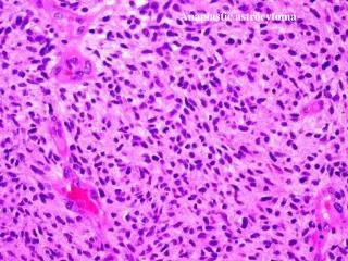
Supplemental Figure1. Histological appearance of WHO GradeIII anaplastic meningioma. . B. A. *. C. *. *. Supplemental Figure 1. Histological appearance of WHO Grade III anaplastic meningioma

E N D
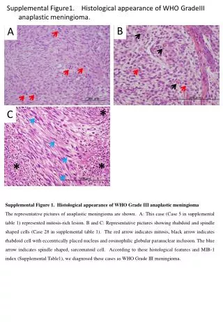
Supplemental Figure1. Histological appearance of WHO GradeIIIanaplastic meningioma. B A * C * * Supplemental Figure 1.Histological appearance of WHO Grade III anaplastic meningioma The representative pictures of anaplastic meningioma are shown. A: This case (Case 5 in supplemental table 1) represented mitosis-rich lesion. B and C: Representative pictures showing rhabdoidand spindle shaped cells (Case 28 in supplemental table 1). The red arrow indicates mitosis, black arrow indicates rhabdoid cell with eccentrically placed nucleus and eosinophilic globular paranuclear inclusion. The blue arrow indicates spindle shaped, sarcomatoid cell. According to these histological features and MIB-1 index (Supplemental Table1), we diagnosed these cases as WHO Grade III meningioma.