Download
1 / 30
430 likes | 1.73k Vues
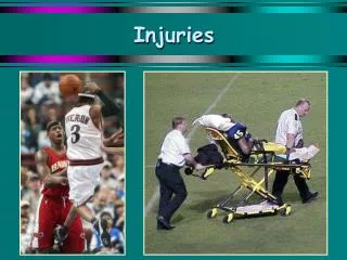
Growth Plate Injuries. Michael LaBella. Objectives. Learn the Anatomy of a growth plate and how it can fracture, as well as who is prone to this type of injury. Identify Key terms associated with Growth Plate Injuries.

E N D
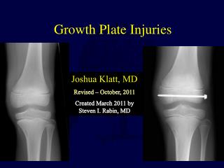
Growth Plate Injuries Michael LaBella
Objectives • Learn the Anatomy of a growth plate and how it can fracture, as well as who is prone to this type of injury. • Identify Key terms associated with Growth Plate Injuries. • Determine the causes and mechanism of injury in the growth plate and distinguish between different types of growth plate injury. • Recognize and evaluate a growth plate fracture or injury. • Suggest treatments and rehabilitation methods for growth plate fracture. • Suggest methods for prevention of growth plate fracture.
Key Terminology Associated with Growth Plate Fractures • Displaced fracture - A fracture in which the two ends of the broken bone are separated from one another. • Epiphysis - The end of a long bone, which is initially separated by cartilage from the shaft of the bone and develops separately. It eventually fuses with the shaft (diaphysis) of the bone to form a complete bone. • Epiphyseal plate - The area of developing tissue near the end of the long bones in children and adolescents. It is also called the physis or growth plate. • Growth plate - The area of developing tissue near the end of the long bones in children and adolescents. Each long bone has at least two growth plates: one at each end. The growth plate determines the future length and shape of the mature bone. When growth is complete - sometime during adolescence - the growth plates are replaced by solid bone. The growth plate is also called the physis or epiphyseal plate. • Metaphysis - The growing portion of a long bone that lies between the ends of the bones (epiphyses) and the shaft (diaphysis). • Physis - The area of developing tissue near the end of the long bones in children and adolescents. The physis is also called the growth plate.
Anatomy- What is the Growth Plate? • The growth plate, also known as the epiphyseal plate or physis, is the area of growing tissue near the ends of the long bones in children and adolescents. Each long bone has at least two growth plates: one at each end. The growth plate determines the future length and shape of the mature bone. When growth is complete - sometime during adolescence - the growth plates close and are replaced by solid bone. • Because the growth plates are the weakest areas of the growing skeleton - even weaker than the nearby ligaments and tendons that connect bones to other bones and muscles - they are vulnerable to injury. Injuries to the growth plate are called fractures.
Anatomy: Who Gets Growth Plate Fractures? • Growth plate injuries can occur in growing children and adolescents. In a child, a serious injury to a joint is more likely to damage a growth plate than the ligaments that stabilize the joint. Trauma that would cause a sprain in an adult might cause a growth plate fracture in a child. • Growth plate fractures occur twice as often in boys as in girls, because girls’ bodies mature at an earlier age than boys. As a result, their bones finish growing sooner, and their growth plates are replaced by stronger, solid bone. • One-third of all growth plate injuries occur in competitive sports such as football, basketball, or gymnastics, while about 20 percent of growth plate fractures occur as a result of recreational activities such as biking, sledding, skiing, or skateboarding. • Fractures can result from a single traumatic event, such as a fall or automobile accident, or from chronic stress and overuse. Most growth plate fractures occur in the long bones of the fingers (phalanges) and the outer bone of the forearm (radius). They are also common in the lower bones of the leg (the tibia and fibula).
Growth Plate Injuries Classified by the Salter-Harris System
Salter-Harris Fracture: Type I The epiphysis is completely separated from the end of the bone, or the metaphysis. The vital portions of the growth plate remain attached to the epiphysis. Only rarely will the doctor have to put the fracture back into place, but all type I injuries generally require a cast to keep the fracture in place as it heals. Unless there is damage to the blood supply, the likelihood that the bone will grow normally is excellent.
Salter-Harris Fracture: Type I Characteristics: Displaced, or widened at the physis. In the radiograph on the right, the epiphysis of the radius is displaced (to the right) at the physis in comparison to the metaphysis. A fracture is not visible in either the epiphysis or the metaphysis. These injuries are associated with a favorable prognosis regardless of region.
Salter-Harris Fracture: Type II This is the most common type of growth plate fracture. The epiphysis, together with the growth plate, is partially separated from the metaphysis, which is cracked. Unlike type I fractures, type II fractures typically have to be put back into place and immobilized for normal growth to continue. Because these fractures usually return to their normal shape during growth, sometimes the doctor does not have to manipulate this fracture back into position.
Salter-Harris Fracture: Type II Characteristics: Fracture through the physis and metaphysis. The plain-film shows a type II Salter-Harris fracture at the distal end of the radius (along with a fracture at the distal end of the ulna). The fracture line passes through the metaphysis into the epiphyseal plate, but no fracture is observed into the epiphysis. These injuries may produce minimal shortening; rarely do these injuries result in functional limitations, except at the knee and ankle.
Salter-Harris Fracture: Type III This fracture occurs only rarely, usually at the lower end of the tibia, one of the long bones of the lower leg. It happens when a fracture runs completely through the epiphysis and separates part of the epiphysis and growth plate from the metaphysis. Surgery is sometimes necessary to restore the joint surface to normal. The outlook or prognosis for growth is good if the blood supply to the separated portion of the epiphysis is still intact, if the fracture is not displaced, and if a bridge of new bone has not formed at the site of the fracture.
Salter-Harris Fracture: Type III Characteristics: Fracture through the physis and the epiphysis. The radiograph displays a fracture of the distal tibia involving the epiphyseal plate, passing through the epiphysis into the articular surface. The physis is widened at the lateral aspect and the medial aspect is closed. This type is prone to chronic disability because it typically involves the articular surface of the joint, but generally has a favorable prognosis.
Salter-Harris Fracture: Type IV This fracture runs through the epiphysis, across the growth plate, and into the metaphysis. Surgery is needed to restore the joint surface to normal and to perfectly align the growth plate. Unless perfect alignment is achieved and maintained during healing, prognosis for growth is poor. This injury occurs most commonly at the end of the humerus (the upper arm bone) near the elbow.
Salter-Harris Fracture: Type IV Characteristics: Fracture through the metaphysis, physis, and epiphysis. The radiograph on the right shows two bone fragments at the medial aspect of the distal tibia--one at the metaphysis, the other at the epiphysis. The fracture passes through the epiphyseal plate, so this demonstrates a type IV Salter-Harris fracture. These injuries can produce joint deformity with angulation more likely at the knee and ankle.
Salter-Harris Fracture: Type V This uncommon injury occurs when the end of the bone is crushed and the growth plate is compressed. It is most likely to occur at the knee or ankle. Prognosis is poor, since premature stunting of growth is almost inevitable.
Salter-Harris Fracture: Type V Characteristics: Compression or crush injury of the epiphyseal plate with no associated epiphyseal or metaphyseal fracture. • Initial plain film x-rays are normal. The diagnosis is more often made in retrospect as premature closing is observed in a physis previously considered uninjured. • The mechanism of injury is also a key factor in making this diagnosis--resulting from compression. • These injuries have a poor prognosis because angulation and/or shortening are 100%.
Salter-Harris Fracture: Type VI A newer classification, called the Peterson classification, adds a type VI fracture, in which a portion of the epiphysis, growth plate, and metaphysis is missing. This usually occurs with an open wound or compound fracture, often involving lawnmowers, farm machinery, snowmobiles, or gunshot wounds. All type VI fractures require surgery, and most will require later reconstructive or corrective surgery. Bone growth is almost always stunted.
Mechanism of Injury- What Causes Growth Plate Injuries? • Growth plate injuries can be caused by an event such as a fall or blow to the limb, or they can result from overuse. For example, a gymnast who practices for hours on the uneven bars, a long-distance runner, and a baseball pitcher perfecting his curve ball can all have growth plate injuries.
Mechanism of Injury Continued • Although many growth plate injuries are caused by accidents that occur during play or athletic activity, growth plates are also susceptible to other disorders, such as bone infection, that can alter their normal growth and development. Other possible causes of growth plate injuries include the following: • Child abuse - More than 1 million children each year are the victims of substantiated child abuse or neglect. The second most common injury among abused children is a fracture, and growth plate injuries are prevalent because the growth plate is the weakest part of the bone. • Injury from extreme cold (for example, frostbite) - Exposure to extreme cold can damage the growth plate in children and result in short, stubby fingers or premature degenerative arthritis (breakdown of the joint cartilage). • Radiation and medications - Research has suggested that chemotherapy given for childhood cancers may negatively affect bone growth. Prolonged use of steroids for inflammatory conditions such as juvenile idiopathic arthritis can also harm bone growth.
Mechanism of Injury Continued • Neurological disorders - Children with certain neurological disorders are prone to growth plate fractures, especially at the ankle and knee. Children who are born with insensitivity to pain can have similar types of injuries. • Genetics - The growth plates are where many inherited disorders that affect the musculoskeletal system appear. Scientists are just beginning to understand the genes and gene mutations involved in skeletal formation, growth, and development. This new information is raising hopes for improving treatment for children who are born with poorly formed or improperly functioning growth plates. • Metabolic disease - Disease states such as kidney failure and hormone disorders can affect the growth plates and their function. The bone growth of children with long-term conditions of this kind may be negatively affected.
Evaluation • Signs that coaches should look for on the field that may point towards a Growth Plate Fracture: • Inability to continue play because of pain following an acute or sudden injury. • Decreased ability to play over the long term because of persistent pain following a previous injury. • Visible deformity of the child's arms or legs. • Severe pain from acute injuries that prevent the use of an arm or leg.
Recognition/Diagnosis of Injury A child who has persistent pain, or pain that affects athletic performance or the ability to move and put pressure on a limb, should never be allowed or expected to “work through the pain.” Whether an injury is acute or due to overuse, it should be evaluated by a doctor, because some injuries, if left untreated, can cause permanent damage and interfere with proper growth of the involved limb. • The doctor will begin the diagnostic process by asking about the injury and how it occurred and by examining the child. The doctor will then use x rays to determine if there is a fracture, and if so, the type of fracture. Often the doctor will x ray not only the injured limb but the opposite limb as well. • Because growth plates have not yet hardened into solid bone, neither the structures themselves nor injuries to them show up on x rays. Instead, growth plates appear as gaps between the shaft of a long bone, called the metaphysis, and the end of the bone, called the epiphysis. • By comparing x rays of the injured limb to those of the noninjured limb, doctors can look for differences that indicate an injury.
Recognition of Injury Continued • Very often an x ray is negative, because the growth plate line is already there, and the fracture is undisplaced (the two ends of the broken bone are not separated). The doctor can still diagnose a growth plate fracture on clinical grounds because of tenderness of the plate. Children do get ligament strains if their growth plates are open, and they often have undisplaced growth plate fractures. • Other tests doctors may use to diagnose a growth plate injury include magnetic resonance imaging (MRI), computed tomography (CT), and ultrasound. • Because these tests enable doctors to see the growth plate and areas of other soft tissue, they can be useful not only in detecting the presence of an injury, but also in determining the type and extent of the injury.
Treatment • Treatment for growth plate injuries depends on the type of injury. In all cases, treatment should be started as soon as possible after injury and will generally involve a mix of the following: • Immobilization • The affected limb is often put in a cast or splint, and the child is told to limit any activity that puts pressure on the injured area. • Manipulation or Surgery • If the fracture is displaced (meaning the ends of the injured bones no longer meet as they should), the doctor will have to put the bones or joints back in their correct positions, either by using his or her hands (called manipulation) or by performing surgery. Sometimes the doctor needs to fix the break and hold the growth plate in place with screws or wire. After the procedure, the bone will be set in place (immobilized) so it can heal without moving. This is usually done with a cast that encloses the injured growth plate and the joints on both sides of it. The cast is left in place until the injury heals, which can take anywhere from a few weeks to 2 or more months for serious injuries. The need for manipulation or surgery depends on the location and extent of the injury, its effect on nearby nerves and blood vessels, and the child’s age.
Treatment Continued • Strengthening and Range-of-Motion Exercises • These are exercises designed to strengthen the muscles that support the injured area of the bone and to improve or maintain the joint’s ability to move in the way that it should. Your child’s doctor may recommend these after the fracture has healed. A physical therapist can work with your child and his or her doctor to design an appropriate exercise plan. • Long-Term Follow-up • Long-term follow-up is usually necessary to monitor the child’s recuperation and growth. Evaluation includes x rays of matching limbs at 3- to 6-month intervals for at least 2 years. Some fractures require periodic evaluations until the child’s bones have finished growing. Sometimes a growth arrest line (a line on the x ray where the bone stopped growing temporarily) may appear as a marker of the injury. Continued bone growth away from that line may mean there will not be a long-term problem, and the doctor may decide to stop following the patient.
Rehabilitation • About 85 percent of growth plate fractures heal without any lasting effect. Whether an arrest of growth occurs depends on the treatment provided, and the following factors, in descending order of importance: • Severity of the injury - If the injury causes the blood supply to the epiphysis to be cut off, growth can be stunted. If the growth plate is shifted, shattered, or crushed, the growth plate may close prematurely, forming a bony bridge or “bar.” The risk of growth arrest is higher in this setting. An open injury in which the skin is broken carries the risk of infection, which could destroy the growth plate. • Age of the child - In a younger child, the bones have a great deal of growing to do; therefore, growth arrest can be more serious, and closer surveillance is needed. It is also true, however, that younger bones have a greater ability to heal. • Which growth plate is injured - Some growth plates, such as those in the region of the knee, are more involved in extensive bone growth than others. • Type of fracture - Of the 6 fracture types described earlier, types IV, V, and VI are the most serious.
Rehabilitation Continued • Regular follow-up visits to the doctor should continue for at least a year after the fracture. Complicated fractures (types IV, V, and VI) as well as fractures to the thighbone (femur) and shinbone (tibia) may need to be followed until the child reaches skeletal maturity. • Physical therapy focuses on more dynamic and functional training to ensure that the patient is able to safely return to his or her previous lifestyle. • For athletes, sport-specific training must be incorporated and the physical therapist must evaluate the overall progression of the healing of the injury in order to provide recommendations to the physician and patient.
Return to Play • Each athlete is treated on an individual basis. The athlete should be off all pain medications, be relatively pain free, and have no return of symptoms during sports-specific activities in order to return to the sport. • Length of time needed for recovery for injury may be sports specific, and can be different depending on position played in a specific sport.
Prevention • Decide if a child is ready for team sports. The American Academy of Pediatrics recommends team sports only for children six years of age and older. • Mental and emotional ability: most children younger than six don't understand the concept and rules of team play, and may not have the emotional development and eagerness to play. • Check the gear. Equipment should be age appropriate and fit correctly. Worn items should be replaced. • Teach children not to play through pain. It won’t make him tougher and it could cause her to make an injury even worse. • Warm up and stretch. Warm up and cool down exercises, such as stretching and light jogging, can help minimize the chance of muscle strain or other soft tissue injuries during sports. • Let injuries heal completely. When a growth plate has been injured, minimize long-term damage by allowing the affected area to heal completely before participating in the sport again.
References American Academy of Orthopaedic Surgeons (2008). Growth Plate Fractures. Publication Available Online: http://orthoinfo.aaos.org American Orthopaedic Society for Sports Medicine. (2008). Publication Available Online: http://www.sportsmed.org/tabs/Index.aspx Caine, d, DiFiori,J., Maffulli,N. (2006). Physeal injuries in children's and youth sports: reasons for concern?British Journal of Sports Medicine (40), (9), 749-760. National Institute of Arthritis and Musculoskeletal and Skin Diseases (NIAMS)Information Clearinghouse. National Institute of Health (2007). Growth Plate Injuries. Publication Available Online: http://www.nih.gov