Cardiac ECMO
EACTS/ Hannes Meyer symposium 2012. Cardiac ECMO. CJ Jordaan Dept. Cardiothoracic surgery and Critical care University of the Free state Bloemfontein. First SUCCESFULL case in 1976 ECMO had several limitations Due to poor outcomes (poor indications )

Cardiac ECMO
E N D
Presentation Transcript
EACTS/ Hannes Meyer symposium 2012 Cardiac ECMO CJ Jordaan Dept. Cardiothoracic surgery and Critical care University of the Free state Bloemfontein
First SUCCESFULL case in 1976 • ECMO had several limitations • Due to poor outcomes (poor indications) • ECMO had high mortalities worldwide • High costs involved • Only few selected centres worldwide persisting • THEN CAME THE REBIRTH OF ECMO
Cardiac (V-A) ECMO support: Indications “Any patient may benefit from ECMO if they have severe respiratory or cardiac failure or both that is refractory to conventional treatment, providing that the underlying disease is ‘potentially reversible’ and there is no absolute contra-indication for support“ ‘potentially reversible’ : Post surgery for congenital heart disease …technically satisfactory repair Bridge to transplant: Availability of donor organs ELSO guidelines, 2011
ECMO indications CARDIAC: • Post cardiotomy • Transient myocardial dysfunction • Transiently increased PVR • Stabilisation of cardiac patients preoperatively • Arrhythmia • Bridge to transplant • Non heart beating organ donation
ECMO indications.. Respiratory: • ARDS • Pneumonia • Trauma • Primary graft failure • Paediatric population: • Meconium aspiration • Congenital diaphragm herniation • Cardiac support • Pulmonary hypertension
Cardiac (V-A) ECMO support: Indications University of Michigan Criteria Cardiac index < 2 l/m2 per min for 3 h Metabolic acidosis Base deficit > 5 for 3 h Blood Pressure Neonate MAP < 40 mm Hg Infant MAP < 50 mm Hg Child MAP < 60 mm Hg Urine < 0.5 ml/hr After an operation Technically satisfactory repair and failure to wean off bypass
Cardiac (V-A) ECMO support: Contra-indications Actual or possible major brain injury Prolonged shock Low Blood Pressure Metabolic acidosis > 5 for 12 h Oliguria <0.5 ml/h for >12 h After an operation Technically unsatisfactory repair Disseminated malignancy Advanced age Unwitnessed cardiac arrest Prolonged resuscitation Aortic insufficiency
Neonatal criteria. • Gestational age ≥ 34 weeks or • Birth weight ≥ 2000g • No evidence of significant coagulopathy or uncontrolled bleeding • No major intracranial haemorrhage, < grade 2 IVH • Reversible lung disease with length of mechanical ventilation < 10-14 days • No correctable congenital heart disease • No lethal congenital anomalies • No evidence of irreversible brain damage
Respiratory criteria: • AaDO > 605-620 mmHg for 4- 12 hours • Oxygenation index > 35-60 for 1– 6 hours • PaO₂ < 35- 60 mmHg 2-12 hours • pH < 7,25 for 2 hours or with hypotension • Acute deterioration
Pump head: • Magnetic levitation of the impeller • No wear resulting in reduced downtime and low maintenance cost • No lubrication required plus the capability of running dry • No particle generation • Wide clearances between the pump casing and impeller, and therefore, no clotting or stopping of the pump • Simple cleaning, sterilization, and exchange of the pump casing and/or impeller • Wide temperature range • Extremely low vibration and noise
PMP Oxygenators • Diffusion membrane made from polymethylpentene (PMP). • Woven into a complex configuration of hollow fibers. • low resistance configuration mat arranged in well defined stacks • maximum blood/gas mixing. • Gas transfer takes place without the direct contact with blood. • PMP membrane surface in contact with blood is treated with a heparin coating to provide a biocompatible and non-thrombogenic surface. • Blood flows over the exterior surface of the device’s fibers
Cannula Design - Physics • Poisseulle’s Law Flow αRadius4 x Pressure Length • In other words short and fat is best
Management • ECMO • Flow and FiO2 governs oxygen delivery • Sweep governs CO2 clearance • VA flow adjusted by monitoring MVO2 and lactate • Target value > 75% • Beware L R Shunt
Management • Ventilation • Low tidal volume ventilation • Relatively little blood through the lungs • Coronary perfusion • Occasionally HFOV • Nitric only for trial off ECMO • Ventilator settings: • TV 5-6 ml/kg • PEEP 5-10 cmH20 • PIP 20 cmH2O • RR 10 b/m • FiO2 <100%
Inotropes • Usually weaned after cannulation • Some background inotropic support beneficial to maintain contractility • IABP may also benefit • Problem with LV distension
Management • Fluid balance • Try to keep patient as dry as possible • Aggressive diuretic therapy • Early CVVH
Management • Echocardiography • Monitor cardiac function • Distension of heart • Consider venting or septostomy • Early Cardiac Catheter to aid decisions • Drop in ECMO flow: • Volume dependent • Cannula malposition • Pneumothorax or tamponade • Increased afterload
The accurate and secure location of adequately sized cannula is a pre-requisite to successful ECMO • Techniques vary depending • Mode of ECMO • Age of patient
Imaging • Ultrasound • Guide percutaneous placement • Sizing of vessel • Conformation of position • Fluroscopy • Guidewire and cannula placement
Paediatric and Adults • Percutaneous Technique • Number, size and position of cannula depends on desired flow • Vessels used • Right internal jugular • Right femoral • Left femoral • Rarely left internal jugular • HEPARIN GIVEN • Guidewire inserted • Progressive dilitation • Cannula inserted and secured
Double lumen cannulation of right internal jugular vein • Patient prepped and draped • Transverse cervical incision • Diathermy to prevent subsequent haemorrhage • Divide platysma • Retract sternomastoid laterally • Division of omohyoid may improve access
3 Cannula reduce recirculation Femoral Femoral cannulation may give up to 60% re-circulation Drainage from neck may decrease cerebral venous hypertension
New cannula – Avalon Elite • Improved handling • Kink and collapse resistant • Reduced re-circulation in sheep to around 2% • Potential for adult VV support through single double lumen cannula • Sizes available • 13, 16, 19, 20, 23, 27 and 31 Fr available
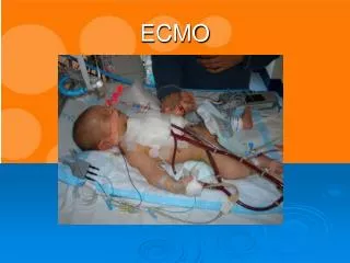
VA Cannulation • Open technique although percutaneous possible • Peripheral or trans-thoracic cannulation • Age and size dependent variation as with VV cannulation Central ECMO cannulation Peripheral ECMO cannulation
Carotid and right internal jugular vein exposed • HEPARIN GIVEN • Vessel ligated cranially • Vascular clamp applied proximally • Arrowhead incision • Clamp removed as cannula slipped into vessel • Cannula tied in proximally • Process repeated for venous cannula
Distal perfusion • Important consideration when end arteries cannulated • Small return cannula • Y shaped cannula • Distal cannulation • Vessel sparing cannulation
VA Trans-thoracic Cannulation • Often used for failure to wean from bypass • May provide rapid cannulation in ECPR • Drainage right atrium • Return aortic root • Avoids carotid sacrifice
VA Trans-thoracic Cannulation • Advantages • Requires minimal additional dissection • Good drainage/return • Improved cerebral perfusion • Increased venting options • Disadvantages • Cannula more likely to dislodge • Increased bleeding • Decreased respiratory function with open chest • Infection risk
Peripheral VA Cannulation • Femoral vessels too small in non-walkers • Carotids commonly used • Venous drainage from right internal jugular vein or femoral vein • Semi-seldinger technique may reduce local vessel damage aiding decannulation
Management • Bleeding • Be prepared to pack chest and re-explore • Switch to peripheral cannulation • Close chest • Stepwise approach • Dipyramidole • Aspirin • Aprotinine • Antifibrinolytics