Case
This case study discusses a patient with 1st metatarsophalangeal joint pain, radiologic findings, MRI results, surgical excision, and pathologic diagnosis of calcium pyrophosphate dihydrate crystal deposition disease. The etiology, clinical patterns, diagnostic imaging, and management strategies are elaborated. This includes the clinical features, imaging modalities, and treatment options available for this condition.

Case
E N D
Presentation Transcript
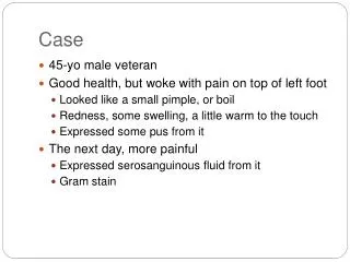
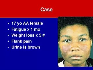
Case • M/23 • C.C. : 1st MTP joint pain (1YA)
Radiologic findings-CT • Expansile osteolytic mass at medial sesamoid of hallux - with suspicious fracture lines -> DDx) 1. chondroblastoma or GCT 2. ABC 3. intraosseous gout • Rec) Rt foot MRI using microcoil
T1 sagittal T1 coronal T1 sagittal (FS/Gd+) 20070220 Foot MRI (Rt.)_Contrast
T1 axial T2 axial T1 axial (FS/Gd+) 20070220 Foot MRI (Rt.)_Contrast
Radiologic findings-MRI • Expansile mass involving medial sesamoid of hallux - peripheral irregular thick enhancement and central nonenhancing cystic or necrotic area - with its associated synovial enhancement of lst MTP joint - with bony erosion at lst metatarsal neck c reactive bone marrow edema • vascular structures anterior to the mass • closely abutting flexor hallucis tendon ->DDx) 1. intraosseous gout,most likely 2. tbc 3. tumorous condition such as giant cell tumor or chondroblastoma
Hospital course • Op: Excision of Sesamoid, Rt.(2007-02-26) • Pathologic diagnosis • Soft tissue, right foot, excision • 1. Numerous rhomboid crystals showing birefringence • 2. Some foci of calcium deposit • 3. Chronic granulomatous inflammation with 1) multinucleated giant cells 2) central hyaline degeneration consistent with calcium pyrophosphate dihydrate deposition disease
Calcium pyrophosphate dihydrate (CPPD) crystal deposition disease • Etiology • Idiopathic: most common • Increased with age (7% of population near age 70 and 30-60% by the age 80) • Hereditary: autosomal dominant condition • Maybe associated with ANK(chromosome 5p15) • Secondary: 5-10% of patients have metabolic disease. • Hyperparathyroidism, Hemochromatosis, Hypophosphatasia… • Clinical patterns • Asymptomatic chondrocalcinosis • CPPD crystal arthropathy • Pseudogout (18%), pseudo-osteoarthritis with/without synovitis (40%/18%), pseudorheumatoid arthritis (8%) • Common location: Knee, wrist, MCP joint Seminars in musculoskeletal radiology 2003;07:175-186
Calcium pyrophosphate dihydrate (CPPD) crystal deposition disease • Diagnositic imagings • Conventional radiography • Calcification within or around joints • Chondrocalcinosis • Synovial and capsular calcifications • Other soft tissue calcification • Findings of pyrophosphate arthropathy • Bilateral, symmetrical involvement of affected articulations • Cartilage loss, subchondral plate sclerosis, subchondral cyst formation • Subchondral collpase, fragmentation, intra-articular loose body • MRI • Less dense calcium deposition->GRE sequence is more sensitive than conventional radiography. Seminars in musculoskeletal radiology 2003;07:175-186