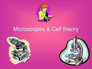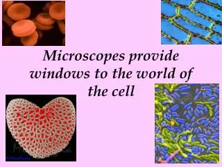
Microscopes and the Develop of the Cell Theory
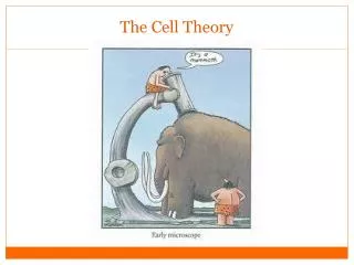
Microscopes and the Develop of the Cell Theory. 1665 Robert Hooke. Used Compound Light Microscope. Find the lenses… Look at the light source!. Aaaahhh ! What is this?. Aaaahhh ! What is this?. 1665 HOOKE – named cells after looking at cork

Microscopes and the Develop of the Cell Theory
E N D
Presentation Transcript
Used Compound Light Microscope Find the lenses…Look at the light source!
1665 HOOKE –namedcells after looking at cork (cell walls) and seeing the resemblance to monk cells
Credited for observing living organisms, or “animalcules”
1838 Matthias SCHLIEDEN 1839 Theodor SCHWANNplants are made of cellsanimalsare made of cells
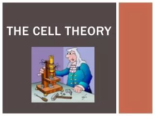
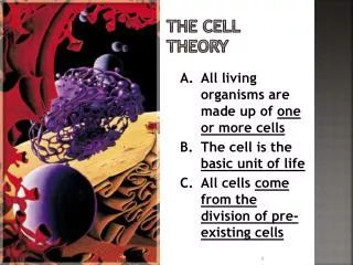
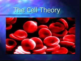
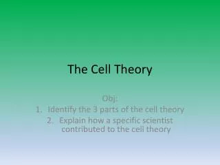
Cell Theory • All organisms are made of cells • Cells are the basic unit of structure & function • Cells come only from preexisting cells
LIGHT MICROSCOPE • Uses light and lenses to directly observe specimens • Compound: 2 lenses, multiply lens powers • Simple: 1 lens
TEM • Transmitting e- microscope Image magnified more than 100,000’s times Thin slice of specimen Bombarded w/beam of e-s e-s pass through specimen (similar to light/density) Image interpreted by computer
SEM • Scanning e- microscope 3-D image magnified more than 100,000’s times Specimen coated in gold/metal Bombarded w/beam of e-s e-s bounce off of the outside of the specimen (RADAR) Image interpreted by computer
http://www.sgclub.com/humour/insects-under-the-electron-microscope/http://www.sgclub.com/humour/insects-under-the-electron-microscope/