Elbow Orthopaedic Tests
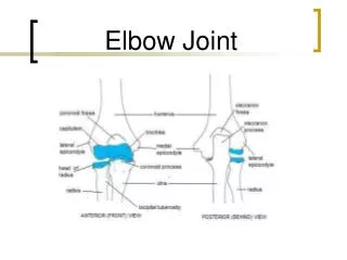
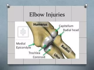
Elbow Orthopaedic Tests. Medial Aspect (Ulnar Nerve). Medial Epicondyle. Ulnar Collateral Ligament. Lateral Epicondyle. Radial Collateral Ligament Annular Ligament. Olecranon Process and Bursa. Triceps. Lateral Epicondylitis (Tennis Elbow).

Elbow Orthopaedic Tests
E N D
Presentation Transcript
Lateral Epicondylitis(Tennis Elbow) • Lateral epicondylitis is a repetitive strain injury of the common extensor tendon at thelateral epicondyle of the humerus. • Symptoms persist because of constant traction movement of the wrist and hand.
Lateral Epicondylitis(Tennis Elbow) • Clinical Signs and Symptoms • Local lateral elbow pain • Weakness of the forearm
Cozen’s Test • Procedure: Patient seated. Stabilize forearm. Patient should make a fist and extend it against resistance. • Rationale: The tendons that extend the wrist attach to the lateral epicondyle. Forcing the extended wrist into flexion will exacerbate the pain if the tendons are inflamed.
Mill’s Test • Procedure: Patient seated. Instruct the patient to pronate the arm and flex the wrist. Then, instruct them to supinate against resistance. • Rationale: The supinator tendon is attached to the lateral epicondyle. If pain is elicited, suspect inflammation of the lateral epicondyle.
Medial Epicondylitis(Golfer’s Elbow) • Medial epicondylitis is a repetitive injury of the common flexor tendon at the medial epiconsyle of the humerus. • Symptoms persist due to constant traction and movement of the wrist and hand.
Medial Epicondylitis(Golfer’s Elbow) • Clinical Signs and Symptoms • Local medial elbow pain • Weakness of the forearm
Golfer’s Elbow test • Procedure: Patient seated. Instruct the patient to extend the elbow and supinate the hand. Then, instruct the patient to flex the wrist against resistance. • Rationale: The tendons that flex the wrist are attached to the medial epicondyle. If pain is elicited, suspect inflammation of the medial epicondyle.
Ligamentous Instability • Ligamentous instability of the elbow is relatively uncommon. • The injury may be caused by forced elbow hyperextension, forced abduction of the extended arm, or forced adduction of the extended arm.
Ligamentous Instability • Forced adduction will damage the radial collateral ligament. • Forced abduction will damage the ulnar collateral ligament.
Ligamentous Instability • Clinical Signs and Symptoms • Medial or Lateral elbow pain • Local swelling
Adduction Stress Test • Procedure: Patient seated. Stabilize the medial arm and place adduction pressure on the patient’s lateral forearm. • Rationale: Adduction pressure will stress the radial collateral ligament. Gapping and pain indicate radial collateral ligament instability.
Abduction Stress Test • Procedure: Patient seated. Stabilize the lateral arm and place abduction pressure on the medial forearm. • Rationale: Abduction pressure on the medial forearm applies stress to the ulnar collateral ligament. Gapping and pain indicate ulnar collateral ligament instability.
Neuropathy / Compression Syndromes • Neuropathy and compression syndromes of the elbow are peripheral neurological disorders. • They are caused by trauma, overuse, arthritis, and postural considerations.
Neuropathy / Compression Syndromes • Paresthesia and weakness of the forearm and/or hand. • The ulnar nerve is most often affected. • Compression occurs in the groove between the olecranon process and the medial epicondyle or the cubital tunnel.
Neuropathy / Compression Syndromes • Clinical Signs and Symptoms • Forearm and/or hand paresthesia • Forearm and/or hand weakness
Tinel’s Sign • Procedure: Patient seated. Tap the ulnar nerve in the groove between the olecranon process and the medial epicondyle with a neurological reflex hammer. • Rationale: If pain is elicited, it suggests a neuritis or neuroma of the ulnar nerve.
Causes of Ulnar Nerve Damage • Excessive use or repetitive motion injuries. • Arthritis of the elbow joint. • Cubital tunnel compression, between the heads of the flexor carpi ulnaris muscle. • Postural habits that compress the nerve, such as sleeping with elbows flexed and hands under head. • Recurrent nerve subluxations or dislocations.