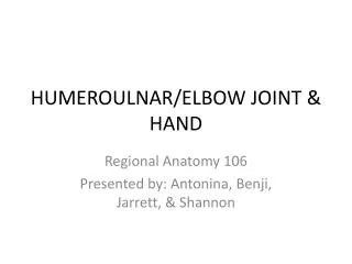
Elbow Joint
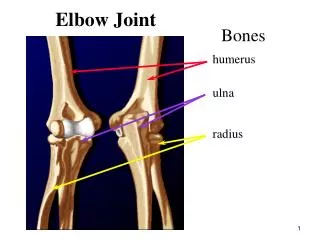
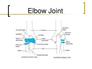
Elbow Joint. Elbow Anatomy. Elbow joint is made of 3 bones 2 joints One capsule Hinge joint Flexion(145) and extension. Elbow Anatomy. Elbow joint- where the radius and ulna articulate with the humerus. Flexion and extension-hinge joint. Elbow Anatomy.

Elbow Joint
E N D
Presentation Transcript
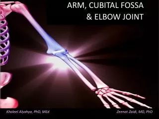
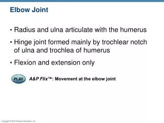
Elbow Anatomy • Elbow joint is made of • 3 bones • 2 joints • One capsule • Hinge joint • Flexion(145) and extension
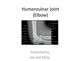
Elbow Anatomy • Elbow joint- where the radius and ulna articulate with the humerus. • Flexion and extension-hinge joint
Elbow Anatomy • Radialulnar joint- where the radius and ulna articulate. • How many of this joint are there???
Elbow Anatomy Radioulnar joint is a pivot joint. It allows supination and pronation.
Elbow Anatomy The ulna does not move. The radius moves around the ulna. The ulna is locked in place by the proximal end at the olecranon process.
Elbow Anatomy Infraglenoid tubercle- inferior lip of the glenoid fossa, where the long head of triceps attaches.
Elbow Anatomy Supraglenoid tubercle- superior portion of the glenoid fossa. Where the long head of biceps attaches.
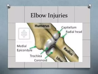
Elbow Anatomy Capitulum- lateral side of joint, it articulates with the radius.
Elbow Anatomy Medial epicondyle- medial side of humerus at distal end just above trochlea. Pronator teres attachment.
Elbow Anatomy Lateral epicondyle- located on lateral side of distal end above capitulum. Anconeus and supinator attach.
Elbow Anatomy • Olecranon fossa- posterior surface of the humerus between the medial and lateral epicondyles.
Elbow Anatomy • Olecranon process- proximal end of ulna, on posterior surface. The point of elbow where triceps attaches.
Elbow Anatomy • Trochlea- located on the medial side of the distal end of the humerus. Articulates with ulna.
Elbow Anatomy • Coronoid Process- just below the trochlear notch and next to the radial notch. Attachment for brachilis.
Elbow Anatomy • Styloid Process- distal end of the lateral/medial surface at the ulna or radius.
Elbow Anatomy • Ulnar head- distal end on the lateral surface, radial head pronates around it.
Elbow Anatomy • Radial head- proximal end, where it articulates with the capitulum.
Elbow Anatomy • Radial tuberosity- attachment for the biceps muscle. On medial side of radius just distal to the radial head.
Elbow Ligaments • Medial Collateral Ligament- runs from medial epicondyle of humerus to the medial side of coranoid process and olecranon process.
Elbow Ligaments • Lateral collateral ligament- attaches proximally to the lateral epicondyle and distally to the lateral ulna and annular ligament.
Elbow Ligaments • Annular Ligament- encompasses radial head at the radial notch and hold it against the ulna. (red)
Elbow • Interosseous membrane- broad flat membrane is located between the radius and ulna for most of their length.
Epicondylitis • Overuse of the tendon attached to the epicondyle of the humerus. • Faulty technique/mechanics, weak muscles or improper equipment. • Can be on lateral side- tennis or golfers elbow • Can be on the medial side- pitcher’s elbow
Signs and Sx Pain over epicondyle Increase pain with wrist flexion or extension Elbow contracture Treatment Proper technique Proper equipment Good warm up with slow increase in intensity Stretching RICE Epicondylitis
Young Athlete’s Elbow Injury • Little Leaguer’s elbow-repetitive action resulting in elbow pain in young • The elbow area is the last epiphyseal center to close so injuries can occur.
Young Athlete’s Elbow Injury • Little Leaguers elbow can result in in varies fractures or bone growths.
Dislocations • Elbow is the second most commonly dislocated major joint. • Most often the ulna/radius dislocate posterior to the humerus. • MOI- fall of outstretched arm with elbow locked in extension.
Signs and Sx Obvious deformity Check circulation and nerve function Treatment Immobilization Wrist strengthening and then progress to elbow RICE Dislocations
Fractures to Elbow/Forarm • Often occurs due to a direct blow or a fall on outstretched arm. • Most common childhood injury, often involves the epiphysis.
Signs and Sx Point tenderness Possible deformity Swelling Limited ROM Treatment Immobilization RICE Strengthening of the joints surrounding and then whole arm when cast is removed. Fractures to Elbow/Forarm Splinting?
Elbow/Forearm Muscles • Brachialis- attaches distal half of the humerus to the coronoid process and ulnar tuberosity of ulna.
Elbow/Forearm Muscles • Biceps Brachii • Long head-supraglenoid tubercle, through bicipital groove, joins with the • Short head- comes from the coracoid process • Both combine to inset onto the radial tuberosity.
Elbow/Forearm Muscles • Brachioradilis- attached to humerus just slightly above the lateral epicondyle, crosses the elbow anterior and lateral to attach on the styloid process of the radius.
Brachialis, Biceps Brachioradialis • Primary Function of these three muscles is what??????
Triceps brachii-entire muscle mass of posterior arm. It attaches to the olecranon process when all 3 heads of the muscle combine. Function is extension. Radial nerve. Elbow/Forearm Muscles
Elbow/Forearm Muscles • Anconeus- helps the triceps with extension and keeps the annular ligament out of the olecranon fossa.
Elbow/Forearm Muscles • Pronator teres- cordlike shape is teres, and pronation is its pimary action. Mostly superficial muscle.
Elbow/Forearm Muscles • Pronator quadratus- small flat quadrilateral muscle that pronates the wrist. Deep muscle at distal end of forearm.
Elbow/Forearm Muscles • Supinator- deep muscle that wraps around the elbow joint laterally from posterior to anterior surfaces.