Download

1 / 39
430 likes | 1.04k Vues
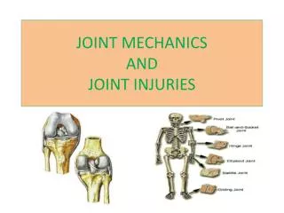
HUMEROULNAR JOINT ( COMMONLY KNOWN AS THE ELBOW JOINT). BRUCE BODDEN & JANELLE JAMES STUDENT PHYSICAL THERAPIST ASSISTANTS. Bones of the Humeroulnar Joint and their fascinating features . Humerus (distal end articulates with ulna and radius):

E N D
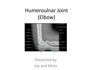
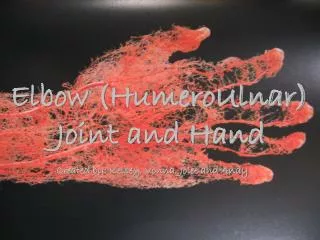
HUMEROULNAR JOINT(COMMONLY KNOWN AS THE ELBOW JOINT) BRUCE BODDEN & JANELLE JAMES STUDENT PHYSICAL THERAPIST ASSISTANTS
Bones of the HumeroulnarJointand their fascinating features Humerus (distal end articulates with ulna and radius): • Capitulum – rounded knoblike surface on lateral aspect • Trochlea – spool-shaped surface located medial to the capitulum • Radial fossa – depression just proximal to the capitulum • Coronoid fossa – depression just proximal to the trochlea • Olecranon fossa – large depression on posterior aspect • Medial epicondyle – pronounced projection on medial aspect • Lateral epicondyle – less prominent projection on lateral aspect
Ulna • Long bone on medial side of forearm – articulates with the trochlea of the humerus • Only bone whose head is distal (at wrist joint, not elbow) • Olecranon – posterior aspect of proximal end: • Hook-shaped process that forms the bony prominence of the elbow • Fits into olecranonfossa of humerus when elbow joint is extended • Coronoid process – anterior aspect of proximal end: • smaller projection that fits into coronoidfossa of humerus when elbow joint is flexed • Trochlear notch – depression between olecranon and coronoid process • Articulates with trochlea of humerus • Radial notch – lateral & inferior to trochlear notch • Articulates with head of radius • Ulnartuberosity– just inferior to coronoid process • Insertion point for biceps brachii
Radius • Long bone on lateral (thumb) side of forearm • Head articulates with the capitulum of the humerus • Neck – just distal to head • Radial tuberosity – just distal to neck. Also an insertion point for biceps brachii • Trochlea of humerus forms a hinge joint with proximal end of ulna (elbow flexion) • Capitulum of humerus and head of radius form a joint that is partly like a hinge, partly ball-and-socket (more on this in a moment) • Head of radius forms a pivot joint with proximal end of ulna (forearm rotation)
Articular capsule • Composed of two layers • Outer fibrous layer – thickened continuation of the periosteum between bones • Dense irregular connective tissue, connects bone to bone • Inner synovial membrane – areolar connective tissue with elastic fibers – lines the internal surface of the fibrous layer • Cells in this layer secrete hyaluronic acid; this combines with interstitial fluid filtered from blood plasma to create synovial fluid, which forms a lubricating film over the articulating surfaces of the joint • All articulating surfaces of the bones in the elbow joint (and most freely-moving joints) are covered with a layer of hyaline articular cartilage which provides a smooth slippery surface to reduce friction between bones. Also helps to absorb shock. • At the sides of the elbow joint, dense irregular connective tissue of the fibrous layer is arranged in parallel bundles called ligaments
Ligaments • Collateral ligaments of the elbow joint • Anular ligament of radius: encircles and holds the head of the radius in the radial notch of the ulna • Forms the proximal radioulnar joint • Permits pronation & supination of forearm • Radial collateral ligament: proximally, extends from lateral epicondyle of humerus; distally, blends with anular ligament of radius • Ulnar collateral ligament: extends from medial epicondyle of humerus to coronoid process & olecranon of ulna. Consists of three bands: • Anterior (cord-like, strongest) • Posterior (fan-like, weakest) • Oblique band (slender; deepens the socket for the trochlea of the humerus) • Interosseous membrane – substantial sheet of dense irregular connective tissue. Binds radius & ulna together and permits slight movement. (There is also one of these connecting the tibia & fibula of the lower leg, but that is not our problem )
Bursae • Pouches of synovial-like fluid located at friction points around joints • Not strictly part of synovial joints, but resemble articular capsules because their walls consist of an outer fibrous membrane of dense connective tissue lined by a synovial membrane • Subcutaneous olecranon bursa: located in the subcutaneous connective tissue over the olecranon • Subtendinousolecranon bursa: located between the olecranon and the triceps tendon (just proximal to its insertion point on the olecranon) • Intratendinous olecranon bursa: sometimes present in the tendon of the triceps brachii
SURFACE ANATOMY • Lateral epicondyle – palpable as a bony point on the outside of the elbow • Medial epicondyle – palpable as a bony point on the inside of the elbow • (since these are part of the humerus they do not move when the elbow flexes or the forearm rotates) • Radial styloid process – palpable as a bony knob at the wrist, just lateral to radial pulse point • Ulnarstyloid process – head of the ulna is palpable (& usually visible) as a raised bony point on the medial (pinky) side of the wrist; styloid process is just distal to that • Olecranon – palpable & visible as the “point” of the elbow during flexion
SURFACE ANATOMY • Cubitalfossa– the hollow on the inside of the elbow • Medial bicipital groove – hollow on the medial side of the upper arm between bellies of the biceps brachii (anterior) and triceps brachii (posterior). Brachial pulse is found here. • Biceps tendon – palpable (& usually visible, with elbow flexion) just proximal to the cubitalfossa. • Triceps tendon – palpable (& often visible) as a ridge on posterior of upper arm, extending proximally from the olecranon • Carrying angle – at full extension, the arm is not a straight line. In anatomical position, the long axis of the ulna makes an angle of ~170o with the long axis of the humerus. • More pronounced in women (~165o) than men (~170o)
Nerves • Radial nerve: originates from posterior cord of brachial plexus • Roots: C5-8, T1 • Motor innervation: triceps brachii and extensors of the hand & wrist • Sensory innervation: most of the back of the hand • Ulnar nerve: originates from medial cord of brachial plexus • Roots: C8 – T1 • Motor innervation: muscles in the forearm that move the hand & wrist (mostly flexors) • Sensory innervation: 5th finger, medial half of 4th finger • Median nerve: originates from lateral & medial cords of brachial plexus • Roots: C5-8, T1 • Motor innervation: pronator teres, pronator quadratus, flexors of hand & wrist • Sensory innervation: parts of palm, some fingertips • Musculocutaneous nerve: originates from lateral cord of brachial plexus • Roots: C5-7 • Motor innervation: biceps brachii, brachialis, coracobrachialis • Sensory innervation: lateral forearm
Arteries • Brachial artery: continuation of subclavian artery ( axillary artery brachial artery) • Supplies blood to nearly all structures of the arm, incl. the humerus • At the cubital fossa, divides to form the radial and ulnar arteries • Deep brachial artery (profunda brachii): arises from brachial artery in upper arm • Supplies posterior compartment of upper arm • Radial artery: arises from brachial artery at cubital fossa (smaller of the two branches) • Ulnar artery: arises from brachial artery at cubital fossa (larger branch)
Arteries, cont’d • Common interosseous artery: arises from ulnar artery in cubital fossa. Gives rise to • Anterior interosseous artery • Posterior interosseous artery • Interosseous recurrent artery: arises from posterior interosseous, between radius & ulna • Superficial palmar arch: direct continuation of ulnar artery • Lies across palm at level of distal border of extended thumb
Veins • Dorsal venous arch: network of veins that drains the dorsal surface of the fingers & hands. Veins come together to form the basilic and cephalic veins. • Basilicvein: travels up the medial side of the arm and joins the brachial vein to become the axillary vein in the mid-arm region • Cephalicvein: travels up the lateral side of the arm and joins the axillary vein in the pectoral region • Median cubital vein: crosses cubitalfossa between basilica & cephalic veins • Median antebrachial vein: drains the venous network of the palmar side of the hand. Ascends the front of the ulnar side of the forearm and ends in the median cubital vein. • Brachial vein: begins at cubitalfossa at the joining of the ulnar & radial veins. Runs parallel to the brachial artery, but deeper. Joins the basilic vein in the axillary region to become the axillary vein.
TENNIS ELBOW CONT… • Tennis elbow is caused by either abrupt or subtle injury of the muscle and tendon area around the outside of the elbow. Tennis elbow specifically involves the area where the muscles and tendons of the forearm attach to the outside bony area (called the lateral epicondyle) of the elbow. Your doctor may call this condition lateral epicondylitis.
CAUSES • microscopic and macroscopic tears between the common extensor tendon and the periosteum of the lateral humeral epicondyle. An operation conducted in this study showed that 28 out of 39 patients showed tearing at the tendon cuff. Radial nerve was significantly involved in tennis elbow: constriction of the radial nerve by adhesions to the capsule of the radiohumeral joint and the short extensor muscle of the wrist.
TREATMENT • Tennis elbow usually is successfully treated by medical means -- such as physical therapy, forearm bracing to rest the tendons, topical anti-inflammatory gels, topical cortisone gels, and cortisone injections. It only rarely requires surgery.