TEMPOROMANDIBULAR JOINT
TEMPOROMANDIBULAR JOINT. ANATOMY PATHOPHYSIOLOGY SURGERY. JOINTS- WHAT ARE THEY?. Human skeleton = endo skeleton It undergoes 2 types of calcification Mesenchyme that becomes calcified is known as intramembranous ossification

TEMPOROMANDIBULAR JOINT
E N D
Presentation Transcript
TEMPOROMANDIBULAR JOINT ANATOMY PATHOPHYSIOLOGY SURGERY
JOINTS- WHAT ARE THEY? • Human skeleton = endoskeleton • It undergoes 2 types of calcification • Mesenchyme that becomes calcified is known as intramembranous ossification • Cartilage that becomes calcified is known as endochondrial ossification • Osseous elements which are multiple that are joined by a variety of structural elements. • These co-aptations are grouped as arthroses • All joints are referred to as an arthroses
ARTHROSES • Stress FORCES • Tensile • Compressive • Shear • Torsion • Tensile forces tend to elongate bones • Compressive forces tend to compress bones • Abnormal forces include shear forces and torsion forces • As it relates to the TMJ, shear forces and torsion forces are produced by parafunctional habits, like nocturnal bruxism and grinding. One concept of ajoint is that it has to be able to move, but not all joints move. • Concerned with differential growth, transmission of forces and with movement
TYPES OF ARTHROSES-2 • SYNARTHROSES: which are solid non-synovial joints. Can be fibrous or cartilaginous. • No synovial membrane • Fibrous denotes intramembranous ossification • Cartilaginous denotes endochondralossification • Abnormal closures of sutures in the cranium will produce abnormal looking faces. Syndromes that could be associated with early closure of synarthroses include Apert’s syndrome, Crouzon’ssyndrom • And in fact these types of syndromes are referred to as the craniosynarthrosessyndromes • Specifically they are an affectation of a synarthroses
TYPES OF ARTHROSES-2 2. DIARTHROSES: which are cavitated synovial joints • Characterized by having articular surfaces covered by cartilage(hyaline or fibrocartilage) with its lubricated smooth wear resistant surface which glides over its fellow with minimal friction • Hyaline cartilage is the most common cartilage associated with articularsurfaces • The TMJ is not composed of with hyaline cartilage, it is fibrocartilage • Cartilage typically resists wear • Within cartilaginous joints there consists a fluid, synovial fluid • Synovial fluid has to be secreted within the joint to allow joint movement • The result of decreased synovial fluid is restricted joint movement
SYNARTHROSES-TYPES • Sutures,synostoses • Gomphoses (peg and socket) • Synchondroses (manubriosternalis) • Remember the face grows downward and forward • Growth sites of the face include the sutures and in particular, in the mandible the growth site is the condyle • So pediatric patients who sustain condyle fractures may have a problem with non-symmetric growth • There are cranial sutures, but there are also fontanelles in the cranium – these are also the site of a synostoses
DIARTHROSES-TYPES • Can be: • Simple(one pair of articulating surfaces) • Compound(more than one pair of surfaces) • Complex(with intracapsular meniscus or disc) • Surface shape: • Plane, spheroid (ball and socket), ellipsoid, ginglymus, bicondylar, trochoid (pivot) and sellar (saddle-shaped) • Example of spheroid shaped joint = hip joint • As it relates to the TMJ, the TMJ is a complex joint and therefore is diarthroidal by definition. • In fact we refer to it as a ginglymoidarthroidal joint
THE TMJ Are the only synovial joints with an articular disc The cartilaginous surfaces are made out of fibrocartilage, not hyaline cartilage
TMJ Classification • Anatomic Classification: • Diarthrodial – two joints (joint spaces) • Superior joint space = above the disc but below the cranium • Inferior joint space = beneath the disc and on top of the condylar head • When you perform joint surgery which joint space do you enter into initally? • Superior • A joint that contains the following characteristics • a freely movable joint • bony surfaces are covered with hyaline or fibrocartilage • lined by a synovial lining and contains synovial fluid Diarthrodial is used because the joint has two articulating bone components – the condyle inferiorly and articular eminence and glenoidfossa superiorly. Ginglymoid is used because the joint has a hinge-like movement component.
TMJ Classification • Functional classification: • Ginglymo– arthroidial = meaning there are two types of movement in the TMJ (this is unusual) • Ginglymoid: rotation - inferior joint space • Arthroidial: translation - superior joint space
Additional Notes: • Injurious forces that affect the joint = shear forces • Shear forces ultimately affect the synovial lining. Once the synovial lining is adversly affect you ultimately see decrease lubrication (a decrease in synovial fluid). • During all movements of the TMJ articulation, it is expected that the TMJ disc stay on top of the condylar head. • When the disc is displaced it is called internal derangement • Internal derangement can be associated with displacement of the disc in an anterior, medial, or lateral component • The most common dislocation is an anterior dislocation • The reason that is so painful, is because the disc has two attachments. It has an anterior attachment and a posterior attachment. When the disc is displaced it is the posterior attachment that causes all of the pain. The reason being, the posterior attachment is referred to as the retrodiscal tissue. It happens to contain all of the nerves of the disc. • Pain associated with internal derangement is usually because of function on the retrodiscal tissue
TMJ Classification • The mandible functions as a class III lever system – VERY IMPORTANT!!!!!!! • Load - occlusal surface • When the lower jaw bone is fractured the whole class III lever system becomes very important. The reason being is at the inferior portion of the mandible we have compressive forces, and as the superior portion (where the teeth are) we have tensile forces. • If the mandible breaks we have a reversal of the force phenomena, when this happens the mandible becomes a very difficult bone to fix in terms of trauma. • Fulcrum - TMJ • Force - muscles of mastication • Includes the masseter, temporalis, and the pterygoids (medial and lateral) • The muscles that are typically involved with TMJ pain and dysfunction are the masseter and the temporalis
Anatomy Overview • Temporomandibular joint… • -the articulation of the mandibular condylar process with the glenoidfossa/articular eminenceaspect of the temporal bone. Also includes the interposed articular disc and collateral ligamentous attachments. • -is an encapsulated, synovial joint. • -classified as a ginglymo-arthroidal joint due to its ability to function as a hinge as well as a gliding type of joint. • Translation is accomplished in the superior joint space • -functions as a Class III lever system.
QUESTION: ANATOMY What are the six (6) components of the temporomandibular joint articulation?
Condyles • The condylar head should be convex anteroposterior and mediolateral • Articular surface of the temporal bone • Capsule • This comes from the zygomatic arch and goes down to the condylar neck • Articulardisc • Made of fibrocartilage & is biconcave • Should be associated with the top of the condylar head with all movement • Ligaments • Lateral pterygoid • This is the specific muscle that is associated with the neck of the condyle
Osseous components • The condylar head has convexities in two dimensions • It has one medial to lateral and one anterior to posterior • So the condylar head should be a rounded structure • A condylar head that may tell you that there is pathology associated with the TMJ articulation may show morphology like flattening of the condylar head, beaking of the condylar head (aka osteophyte formation), erosion of the cartilaginous cap (degenerative joint disease) • When we have problems with the shape of the head of the condyle, almost always you will see wear at the fibrocartilage at the top of the condylarhead • When you have a roughened condylar head you will ultimately have problems with the disc gliding over the head
Osseous Components (Add’t Notes) • The glynoidfossa is in an intimate relationship to the ear, so almost all TMJ pain and dysfunction has auricular discomfort • Usually an individual swallows about 2000 times a day, and you move your jaw joint every time you swallow • We can generate loads up to 750 lb/square inch on the joint when eating • The base of the skull is comprised of the temporal bone, specifically the glenoidfossa and the articular tubercle, also of the temporal bone. • Subluxation is a pathology associated with TMJ articulation. That term means that thecondylar head is anterior to the articular tubercle. • Remember if you have subluxaiton, the reason the patient can’t close their mouth is due to spasticity of the muscles of mastication • To relocate that jaw joint, the appropriate force to apply to the mandible is downward and backward • What would be a surgical remedy to chronic dislocation? • To shave the articular eminence, also referred to as an eminectomy
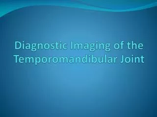
TOMOGRAPHC SCAN OF JOINT • What would be the best imaging modality to look at the disc in TMJ articulation? • MRI • This is a magnet-based technology • It is composed of two different types of images • The T1 image is associated with anatomical detail, so you can see the disc, condylar head, articular eminence, etc • The T2 image is associated with fluid effusions • Typically inflammation induces a fluid effusion • Disc displacement can be diagnosed by an MRI and NOT by a CAT scan • What would be the best imaging modality for a tumor? • You need to know if it is a soft or hard tumor. If you believe it is composed of hard tissue a CAT scan would be best. If you believe it to be soft, an MRI would be best. • Ankylosis may be best diagnosed with a CAT scan
Tomographic Scan of the Joint • When a patient opens his mouth the condylar head should be able to go past or to the articular eminence. • What would you do with a patient who chronically dislocates? • You need to relocated the mandible – this motion would be down and back if the condylar head is in front of the articular eminence • He would then tie the jaws together for 3 weeks • He would then keep her in elastics for 3 weeks to try to retrain her jaw • Another treatment for chronic TMJ dislocation would possibly be eminentically (chop of the eminence)
Bony components • Condyle of mandible • Articular tubercle of temporal bone • Mandibular fossa (glenoid)of temporal bone
SOFT TISSUE COMPONENTS • Muscles and ligaments • Disk • Posterior disk attachment • Joint capsule • Synovia
Elaboration on Soft Tissue Components • Muscles and ligaments • Postural muscles that participate in movement • Paraspinal muscles, trapezius, occipitalis, and sternocleidomastoid • Ligaments associated with articulation • Sphenomandibular ligament & stylomandibular ligament • Disk • Has 4 attachments • Anterior, posterior, medial, and lateral • Posterior disk attachment • Joint capsule –contains the whole joint • Synovia – synovial membrane that contians synovial cells is within the joint space
LIGAMENTOUS SUPPORT • Articular capsule-attached to temporal bone around the edges of the fossa and the articular tubercle and to neck of condyle • This capsule goes anteriorly and surrounds the articular tubercle and to the neck of the condyle • So if you have your condylar process broken secondary to trauma and the condylar segment is displaced you will have disruption of this capsule • Sphenomandibular ligament-medial to the joint, runs from spine of the sphenoid bone to the lingula • There are two rather strong tendinous ligaments, this is the first • The function of this ligament is to maintain the integrity of the nerve as it enters the mandibular foramen, so it is the point of attachment where there is the least amount of movement of the mandible
LIGAMENTOUS SUPPORT CON’T • Stylomandibular ligament-posterior to the joint, runs from the tip of the styloid process to the angle of the mandible • The second tendinous attachment • What happens if the stylomandibular ligament becomes calcified? • Eagle’s Syndrome – painful condition associated with turning of the neck • Temporomandibular ligament-runs from the lateral zygomatic process, anterior to the capsule, to the mandibular neck • A lax TMJ ligament is almost always seen with chronic subluxation
Muscle of Mastication:MASSETER • Location: completely covers the ramus, arises from zygomatic arch and inserts to lateral ramus,deep to the external lobe of parotid gland. It is an elevator of the jaw • Deep to the external lobe of the parotid gland • Function: elevator of the jaw • It is subject to hypertrophy, so hyperactivity (ieparafunctional habits, like clenching, bruxism, and repetitive motions, like gum chewing) all can cause hypertrophy
Muscles of Mastication: Temporalis • Location: arises from the lateral aspect of the cranium, passes deep to the zygomatic arch, and converge onto the coronoidprocess • One problem we see is individuals who have significant parafunctional activity on occasion this temporal muscle extends or enlarges the coronoid process, then this will inhibit maximum incisal opening • So the treatment associated with coronoid process elongation is a coronoidectomy (where you cut the coronoid off) • The problem with doing this is, remember the masseteric artery sits in the coronoid or mandibular notch. When we cut the coronoid process off we typically take the bone out, we don’t let the temporalis retract it back up under the zygomatic arch. • Function: • Anterior and medial fascicles elevate the jaw • Posterior fascicles retract the jaw
Muscles of Mastication: LATERAL PTERYGOID • Location: superior head arises from the infratemporal surface of the sphenoid bone and inserts into the articular capsule and disc; the inferior head arises from the lateral pteryg. pl. and inserts into condylarprocess • So with arthroscopy (when you put a scope into the jaw joint) you can actually see the pterygoid muscle • So if you have disc displacement, by definition, the lateral pterygoid muscle is affected • Function: • Opens the jaw by pulling the condyle and the articular disc anteriorly. • Together, the rt. and lt. lateral pteryg. mm protrude the jaw. • Unilaterally, they swing the jaw to the opposite side and thus effect the grinding motion of mastication • Exursive movements of the lower jaw are produced by action of the lateral pterygoid muscle. The other muscles participate, but left lateral excursions, right lateral excursions, and protrusive excursions are associated with the action of the lateral pterygoid
Muscles of Mastication: MEDIAL PTERYGOID • Deep part arises from the lateral pterygoid plate and the pyramidal process of the palatine bone. The smaller superficial part arises from the tubercle of the maxillla and inserts into the periosteum of the angle of the mandible. Is a strong elevator of the mandible • The medial pterygoid muscle in conjunction with the masseter muscle forms the pterygomasseteric sling • You need to know this so that if you have to place an artificial joint you must disrupt this sling • Function: It is a strong elevator of the mandible
Innervation • Innervation of the muscles of mastication are subserved by the 3rd division of the trigeminal nerve • So the pain associated is mediated by this also • Sensation to the joint itself is provided by two nerves • The primary sensory nerve of the joint if the auriculotemporal nerve • This, of course, is a branch of the mandibular nerve. Remember the mandibular nerve is both sensory and motor. The motor innervation is by the anterior division of the mandibular nerve. The posterior division of the mandibular nerve contains the auriculotemporal nerve. • Secondary innervation is provided by the masseteric nerve
Blood Supply • Blood supply of the TMJ is provided by two arteries: • They are the terminal branches of the external carotid artery • They are the maxillary artery and the superficial temporal artery • He could ask this another way on the exam: Two arteries at risk when performing TMJ surgery are… • Remember, the superficial temporal artery is in the preauricular area and the maxillary artery is medial to the neck of the condyle
Disc Position • Joint noises include pops, clicks, grinding, and crepitus. • Crepitus is the worst joint noise, it is generated by bone on bone • The disc when out of position can be injured • It can be frayed and when the disc has been frayed the articular surface and the lubricating cells have been injured • If you have impingement on the retrodiscal tissue, you have significant pain with mastication • You will see individuals make the claim that they can make a splint that will recapture the disc. Whether that is true or not the real questions is how can you keep the disc in its rightful place with function? • Not uncommonly that requires surgery
Disc Position • Anytime you have abnormal disc position, you can have joint noises. A pop or click is usually associated with the disc coming back to the correct position. • Not uncommonly when you have a click you will have both an opening and closing click
Disc Overview • The disc has four attachments: medial, lateral, anterior, and posterior • The disc is made out of fibrocartilage • The disc within the glenoidfossa anatomically produces a superior and inferior joint space • When performing arthroscentesis or when performing arthroscopy the practitioner enters the superior joint space • Related to the disc the neural innervation is located in the retrodiscal attachment • With disc displacement, the reason you have TMJ arthralgia is because the condylar head is riding on the retrodiscal tissue • With that stated, the normal dislocation vector of a disc is anterior
MOVEMENTS • Elevation/depression occurs in the inframeniscal compartment as the condyle rotates on the articular disc • Protrusion/retraction occurs in the suprameniscal compartment as the articular disc glides anteriorly and posteriorly along the temporal bone between the fossa and the articluartubercle • As it relates to these movements, when you have a total joint, by definition what movement would you not have? • People who have a total joint lose the ability to translate
Anatomy Overview • The superior joint space has about 1.2 cc of volume • The inferior joint space has a smaller volume as compared to the superior joint space • We stated that there is one muscle involved with in the joint – the lateral pterygoid muscle • It has two heads – a superior and inferior head • The superior head is attached to the disc • The inferior head is attached to the neck of the condyle • The blood vessels as well as the nerves are contained in the retrodiscal tissue • The reason the disc cannot repair itself if it has a hole in it is because there is not vascularity within the disc proper
TEMPOROMANDIBULAR DISORDERS • A collective term used to describe a number of related conditions that involve the TMJ’s, masticatory muscles, and associated structures; these conditions may present with facial pain, joint noises, limited jaw function, and other symptoms-ear ache, headache, tinnitus, neck/shoulder pain • Just because the disc is out of place doesn’t mean it is pathologic • Just because the patient has internal derangment (anteriorly displaced disc), doesn’t mean they should have surgery to correct it • He treats if there is pain, and doesn’t if there isn’t • What is the typical headache associated with the TMJ? Tension or migraine? • By definition it should be a tension headache • Many people will come in and tell you they have migraines because they believe every headache is a migraine • Ear ache – you may see ear pathology (like a reddened tympanic membrane) • You may put a child on nitrous and within 3-4 minutes they will start screaming from ear pain • This happens because nitrous tends to diffuse into areas. So if there is a little fissure between the temporotympanic bone and goes behind the tympanic membrane causing ear pain
HISTORICAL REVIEW • Costen syndrome 1934 • Consten found the symptoms of ear ache, head ache, and neck and shoulder pain • Temporomandibular joint pain dysfunction syndrome 1955 • Myofascial pain and dysfunction 1969 • Facial arthromyalgia 1974 • Temporomandibular disorders 1983 • Craniomandibular disorders 1993 • Even though there are many names we still don’t have a consensus on how to treat this pain, we can’t even agree on a surgical technique to use
MOST COMMON TMDs • Myofacialpain and dysfunction – by far the most common • A main component of TMJ problems is myofacial pain • Internal derangement – an anteriorly displaced disc • Osteoarthrosis – degenerative disease • Myofacial pain has a lot of components to it, including a psychological component (stress) • Fibromyalgia is a catch all term, many of his TMJ patients also have fibromyalgia. • TMJ pain and dysfunction is also associated with many other pain syndromes
MYOFASCIAL PAIN AND DYSFUNCTION • Refers to a group of poorly defined muscle disorders (eg, fibromyalgia) characterized by diffuse facial pain and episodic limited jaw opening • May result from parafunctional habits and significant relationship to psychophysiologic disorders such as stress or depression • When you treat this the first thing the insurance company will say is that the patient is crazy. • Insurance companies use the TMJ as an exclusion to not provide treatment
INTERNAL DERANGEMENT • Abnormal relationship of the articular disc to the mandibular condyle, fossa,andarticular eminence, interfering with the smooth action of the joint (Dolwick 1983) • Is a localized mechanical fault within the joint • By definition this implies disc displacement • Synonymous with disc displacement
WILKES CLASSIFICATION • STAGE I: TMJ clicking/no pain/no radiographic DJD • STAGE II: +Jt sounds/pain with intermittent locking but no x-ray DJD • STAGE III: As II, but with x-ray DJD • STAGE IV:ADD without reduction/DJD • STAGE V: Disc perf./Advanced DJD
OSTEOARTHROSIS • Is a nanpainful, localized degenerative joint disease that mainly affects bone and articular cartilage. • It is often idiopathic, but predisposing factors such as old age, repetitive trauma (bruxism), abnormal joint posturing, or multiple surgical procedures may be involved. If painful,then referred to as osteoarthritis
EPIDEMIOLOGY • About 60-70% of the population have features of TMDs • About 20-30% report symptoms of TMDs • About 5% of people with TMD symptoms actually seek treatment • The female:male ranges from 3:1 to 9:1