Download
1 / 73
880 likes | 2.57k Vues
TEMPOROMANDIBULAR JOINT. ANATOMY PATHOPHYSIOLOGY SURGERY. Lecture Dates: 3/30/2011, 4/6/2011, 4/13/2011 He didn’t lecture on some of the first slides in this lecture. The later lectures he lectured on several times. Sorry if it gets confusing. . Decision Paradigm for Surgery.

E N D
TEMPOROMANDIBULAR JOINT ANATOMY PATHOPHYSIOLOGY SURGERY Lecture Dates: 3/30/2011, 4/6/2011, 4/13/2011 He didn’t lecture on some of the first slides in this lecture. The later lectures he lectured on several times. Sorry if it gets confusing.
Decision Paradigm for Surgery • Must be based on rational and appropriate analysis of the disease process • One of the problems we have had with TMJ problems is that we don’t have a surgical protocol to define what a pathology is • For example: if a patient has a click or pop we typically don’t do surgery, if they have that plus pain we typically try to treat it • First thing you would do it pharmacologic intervention, like an NSAID, muscle relaxer, and an occlusal appliance/splint • But once a patient still continues to have pain, dysfunction, limitation of opening, and joint noises perhaps a surgical modality might have to be chosen • What surgical modalities do we have? • We can wash a joint out called arthrocentesis • We can look at it and wash it out, called arthroscopy • Or we can do an open joint procedure – we can either reposition the disc or take the disc out. If we take the disc out we may have to do disc replacement. Presently there is no defined material we universally use for disc replacement. • Finally we have the total joint replacement. • The problem we have here is that there isn’t a universal protocol to treat the joint patient. • Those who cannot remember the past are doomed to repeat it • Jack Kent down in New Orleans made a joint out of sialastic, they found that the Kent prosthesis still continues to this day to destroy peoples lives • This undergoes fragmentation when in the body and causes a superlative foreign body reaction • We see with this prosthesis that patients have dehiscence into the middle cranial fossa with the foreign body reaction, it destroys the condyle, temporal bone, and the glenoidfossa. • This was 25 years ago, and they are still taking them out • Now they have a brand new exposition on the TMJ total joint that is attacking the biomed joint on the market
JOINTS- WHAT ARE THEY? • Osseous elements which are multiple that are joined by a variety of structural elements. • These co-aptations are grouped as arthroses
ARTHROSES • FORCES • Tensile,compressive,shear and torsion • Concerned with differential growth, transmission of forces and with movement
TYPES OF ARTHROSES-2 • 1.SYNARTHROSES: which are solid non-synovial joints. Can be fibrous or cartilaginous. • Fibrous denotes intramembranous ossification • Cartilaginous denotes endochondral ossification
TYPES OF ARTHROSES-2 • 2. DIARTHROSES: which are cavitated synovial joints • Characterized by having articular surfaces covered by cartilage(hyaline or fibrocartilage) with its lubricated smooth wear resistant surface which glides over its fellow with minimal friction
SYNARTHROSES-TYPES • Sutures,synostoses • Gomphoses (peg and socket) • Synchondroses (manubriosternalis)
DIARTHROSES-TYPES • Can be simple(one pair of articulating , surfaces), compound(more than one pair of surfaces), or complex(with intracapsular meniscus or disc) • Surface shape:plane, spheroid(ball and socket), ellipsoid, ginglymus, bicondylar,trochoid(pivot) and sellar(saddle-shaped)
THE TMJ Are the only synovial joints with an articular disc
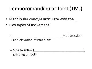
TMJ Classification • Anatomic Classification: • Diarthrodial – two joints (joint spaces) • a joint that contains the following characteristics • a freely movable joint • bony surfaces are covered with hyaline or fibrocartilage • lined by a synovial lining and contains synovial fluid
TMJ Classification • Functional classification: • Ginglymo - arthroidial • Ginglymoid: rotation - inferior joint space • Arthroidial: translation - superior joint space
TMJ Classification • The mandible functions as a class III lever system • Load - occlusal surface • Fulcrum - TMJ • Force - muscles of mastication
Anatomy Overview • Temporomandibular joint… • -the articulation of the mandibular condylar process with the glenoid fossa/articular eminenceaspect of the temporal bone. Also includes the interposed articular disc and collateral ligamentous attachments. • -is an encapsulated, synovial joint. • -classified as a ginglymo-arthroidal joint due to its ability to function as a hinge as well as a gliding type of joint. • -functions as a Class III lever system.
QUESTION: ANATOMY What are the six (6) components of the temporomandibular joint articulation?
Condyles • Articular surface of the temporal bone • Capsule • Articular disc • Ligaments • Lateral pterygoid
Bony components • Condyle of mandible • Articular tubercle of temporal bone • Mandibular fossa (glenoid)of temporal bone
SOFT TISSUE COMPONENTS • Muscles and ligaments • Disk • Posterior disk attachment • Joint capsule • Synovia
LIGAMENTOUS SUPPORT • Articular capsule-attached to temporal bone around the edges of the fossa and the articular tubercle and to neck of condyle • Sphenomandibular ligament-medial to the joint, runs from spine of the sphenoid bone to the lingula
LIGAMENTOUS SUPPORT CON’T • Stylomandibular ligament-posterior to the joint, runs from the tip of the styloid process to the angle of the mandible • Temporomandibular ligament-runs from the lateral zygomatic process, anterior to the capsule, to the mandibular neck
MUSCLES OF MASTICATION • Masseter-completely covers the ramus, arises from zygomatic arch and inserts to lateral ramus,deep to the external lobe of parotid gland. It is an elevator of the jaw • Temporalis-arises from the lateral aspect of the cranium, passes deep to the zygomatic arch, and converge onto the coronoid process
MUSCLES CON’T • Temporalis-anterior and medial fascicles elevate the jaw;posterior fascicles retract the jaw • Lat. Pterygoid-superior head arises from the infratemporal surface of the sphenoid bone and inserts into the articular capsule and disc; the inferior head arises from the lateral pteryg. pl. and inserts into condylar process
MUSCLES CON’T. • Action of Lat. Pteryg.-opens the jaw by pulling the condyle and the articular disc anteriorly. 1)Together, the rt. and lt. lateral pteryg. mm protrude the jaw. 2)Unilaterally, they swing the jaw to the opposite side and thus effect the grinding motion of mastication
MEDIAL PTERYGOID • Deep part arises from the lateral pterygoid plate and the pyramidal process of the palatine bone. The smaller superficial part arises from the tubercle of the maxillla and inserts into the periosteum of the angle of the mandible. Is a strong elevator of the mandible
DISC POSITION • Clicking and popping implies that the disc is out of position • Crepitation implies bone on bone contact
MOVEMENTS • Elevation/depression occurs in the inframeniscal compartment as the condyle rotates on the articular disc • Protrusion/retraction occurs in the suprameniscal compartment as the articular disc glides anteriorly and posteriorly along the temporal bone between the fossa and the articluar tubercle
Anatomy Overview • You must go in through the lateral capsule into the superior disc space. • ML no bigger than 2 cm, AP only 1 cm • If you enter into the jaw joint and accidentally touch the top of the condyle you will wound the fibrocartilage • Fibrocartilage responds to load. The greatest coverage is over the areas of greatest load. • As you start to wear your fibrocartialge down it then produces little pieces of cartilage in the joint. These contribute to adhesion formation.
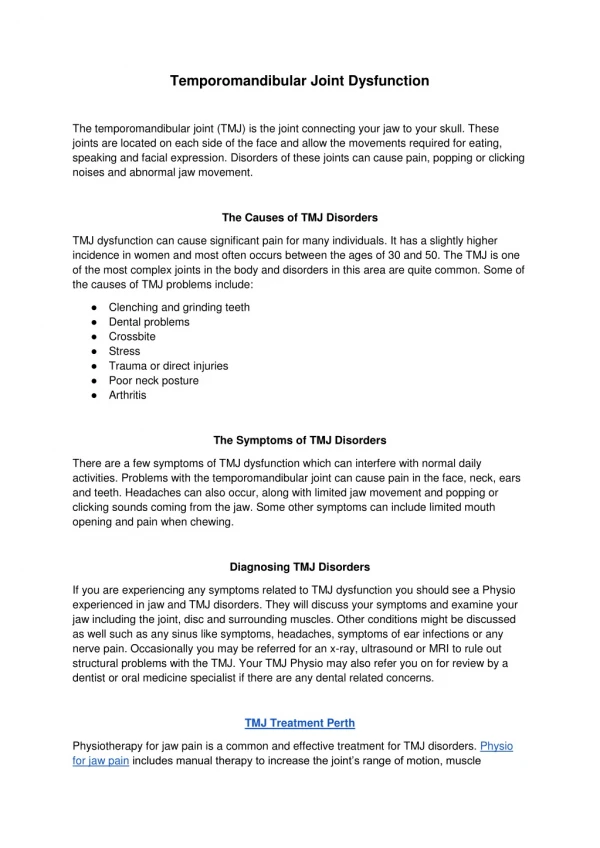
TEMPOROMANDIBULAR DISORDERS • A collective term used to describe a number of related conditions that involve the TMJ’s, masticatory muscles, and associated structures; these conditions may present with facial pain, joint noises, limited jaw function, and other symptoms-ear ache, headache, tinnitus, neck/shoulder pain
HISTORICAL REVIEW • Costen syndrome 1934 • Temporomandibular joint pain dysfunction syndrome 1955 • Myofascial pain and dysfunction 1969 • Facial arthromyalgia 1974 • Temporomandibular disorders 1983 • Craniomandibular disorders 1993
MOST COMMON TMDs • Myofascial pain and dysfunction • You don’t do TMJ surgery for muscle problems. You need to do it either for condyle or disc problems. • Internal derangement • Osteoarthrosis • The problem with the TMJ process is it is complex symptoms so you have complex presentations. When you treat a patient you must be able to get the muscles under control so you can treat the other two parts of the system – the condyle-disc relationship and the teeth
MYOFASCIAL PAIN AND DYSFUNCTION • Refers to a group of poorly defined muscle disorders (eg, fibromyalgia) characterized by diffuse facial pain and episodic limited jaw opening • May result from parafunctional habits and significant relationship to psychophysiologic disorders such as stress or depression
INTERNAL DERANGEMENT • Abnormal relationship of the articular disc to the mandibular condyle, fossa,andarticular eminence, interfering with the smooth action of the joint (Dolwick 1983) • There are two schools of thought on the joint • One believes in arthroscentesis or simple washing out of the joint • The other believes the only way to deal with the jaw joint is through arthroscopy • Is a localized mechanical fault within the joint • Synonymous with disc displacement • Mechanical disturbance of normal function is usually a decrease in maximal incisal opening • Most of the time ID is associated with some type of joint noises, usually a click or pop
Internal Derangement Classification • Most commonly the disc is displaced anteriorly, the disc is usually displaced anteromedially • We do care if the disc is out of place and interferes with function, but they commonly find the disc is out of place postmortem and the patient has had no problems or discomfort with it • Remember there are 4 ligaments of the disc • The reason the disk doesn’t go posteriorly – because the lateral pterygoid is attached to the anterior portion of the disc and the fact that there is no room for the disc posteriorly • He’s never seen it go posteriorly • He has seen the disc displaced superiorly through the glenoidfossa up into the middle cranial fossa due to trauma (MVA)
WILKES CLASSIFICATION • The best classification with the highest success rate is class II • Class II sometimes has a problem with opening and closing their jaw • When you have a disc displacement does this happen typically in the open mouth or closed mouth position? • This typically happens in the closed mouth position. When talking about reduction we are talking about as your open your mouth. • If you get success you will have a success rate of about 70%. Success is defined as a reduction in pain, an increase in function and quality of life. • Locking can occur either in the opening phase or the closing phase. So you can essentially have an open lock or a closed lock. • STAGE I: TMJ clicking/no pain/no radiographic DJD • STAGE II: +Jt sounds/pain with intermittent locking but no x-ray DJD • In stage II there is an instance when the disc doesn’t moved, it called the anchored disc phenomenon. That may be from an adhesion or it may be simply from the dislocation of the disc. The patient will have a reduced maximal incisal opening.
Wilkes Classification cont • STAGE III: As II, but with x-ray DJD • Now you start to see DJD changes • The first DJD one would see would be beaking (the condylar head changes from being nice and round to being kind of pointed, like a birdbeak). Next you would see flattening of the condylar head • This goes through stages. The stages include the initial beaking of the condylar head. You can see subcondyral cyst formation. That is not a real cyst. Subcondryal cyst formation is a thinning in the condylar head and neck region. And then you certainly can have flattening of the condylar head. • One thing to remember about stage II and III is that we still get some disc reduction. When we get to stage IV the disc does not reduce with function. • STAGE IV:ADD without reduction/DJD • STAGE V: Disc perf./Advanced DJD • Is the most complicated stage. This is complicated because the disc has lost its anatomy and now we have a perforation. Now to treat a perforation if you have a central perf of the disc it is not repairable because there is no blood supply there. Peripheral disc perforation can on occasion be sutured together. • Remember the disc is relatively hypovascular, so if you have a hole in it it won’t do any good to try to sew it closed • The vascularity in the disc is provided by the retrodiscal tissues • One problem of course with the disc and trying to get to it is that you have to do a cutaneous approach which brings the facial nerve into play.
Possible Effects of Acute TMJ Trauma • Trauma is the most frequent cause of internal derangement • Other effects of trauma can give us a problem • Ankylosis • Typically seen in patients in prolonged fixation for a condylar fracture • Other causes include: • Frequent middle ear infections • Distraction of the mandible • This causes ankylosis by transporting the condylar segment too fast and too strong. • Distraction is where they take infants who have small mandibles. These infants can’t breathe so they do tracheostomies, this is horrible because they end up with tracheal stenosis. • Instead you place a cut in the mandible and put two pins in the mandible and a bar in it. Every day you crank the mandible and it opens up a little. Over the course of a week it will open up 10 mm. This moves all of the muscles that attach to the mandible with it and helps the patient to breathe without have a tracheostomy. • Part of the problem with this is you push the condyle into the glenoidfossa. After you do this you have to have a period of consolidation where you don’t move the pieces. That is when the patient can get ankylotic. • Intraarticular hemorrhage • Anytime you get a blow to the condyle you can get bleeding into the joint causing fibrosis and TMD • Stretching of ligaments and dislocation • Seen in the whiplash injury, part of what is seen after an MVA. You will need to examine these patients and make a disability rating for them. The TMJ can give you a disability rating of up to 5%. • Tearing of capsule • Can be seen with whiplash injuries • Adhesions restrict the movement of the disc and then cause abnormal joint movement • The problem is when going over the history with the patient they may forget that they have had significant trauma
Single most common antecedent of TMJ internal derangement is trauma
OSTEOARTHROSIS • Is a non-painful, localized degenerative joint disease that mainly affects bone and articular cartilage. • It is often idiopathic, but predisposing factors such as old age, repetitive trauma (bruxism), abnormal joint posturing, or multiple surgical procedures may be involved. If painful, then referred to as osteoarthritis • Predisposing factors: age of patient, repetitive trauma like in bruxism, abnormal joint posture. • Patients who present with class II facial-dental malocclusion (angle’s malocclusion) where you have a relative mandibular hypoplasia, they will try to normalize their appearance. When they do that they project their mandible or push it forward. SO in essence by doing that all day they are almost translating their jaw forward. Overtime that will produce DJD and typically pain in the joint. • It is interesting to note that Class III patients (prognathic mandible) typically don’t have any joint problems. • Another process that will cause osteoarthritis or DJD is multiple surgical procedures. You may have head or people getting cervical steroid injections for neck pain, or knee pain, well you can also do this for TMJ pain. That is in our realm of treatment. So we can place medications within the joint. Remember steroids soften cartilage. So a possible complication is fragmentation of your cartilage. So we say you probably should not give an injection more frequently than once a month, times 3 injections in a year. So one month apart times 3 over one year. Most prudent practitioners however try to inject once every 6 months to prevent this fragmentation process. • If he were to ask on the exam: If a patient has rheumatoid arthritis (a DJD) and it affected both TMJ articulations what would you expect to see clinically? What would you see if a patient has bilateral RA affecting both TMJ’s? • Your condyle-ramus unit shortens on both sides. One would expect to see then premature occlusion of the posterior teeth – so the patient will have an anterior open bite (apertonathia). This is the problem with RA patients. When treating this the RA must burn itself out before doing the surgery or the open bite will recur. To treat this you could reset the maxilla down which will reclose the bite or you can distract the condyle-ramus superiorly. Of those two options, probably the maxillary osteotomy makes more sense, that way you aren’t moving the condylar segment.
DEGENERATIVE CHANGES IN CONDYLE Left: flattening of the condylar head. One can see that as you start into the neck you have real problems and you may have to think about doing a total joint. Right: red = subcondylar cyst, it is a thinning in the condylar head and neck.
EPIDEMIOLOGY • About 60-70% of the population have features of TMDs • About 20-30% report symptoms of TMDs • About 5% of people with TMD symptoms actually seek treatment • The female:male ranges from 3:1 to 9:1
TMJ SURGERY • Indicated for a subset of temporomandibular disorders: There are essentially only 7 reasons to do a surgery 1. Internal derangement – implied disc displacement, typically that disc is displaced anteriorly. 2. Degenerative joint disease – osteoarthritis 3. Rheumatoid arthritis – arthrides, osteoarthritis is a major player in joint pathology. RA is the systemic form of osteoarthritis. The typical presentation when presenting in the joint is the production of apertonathia or open bite. This occurs because you have lost vertical dimension in your condyle-ramus complex. • In addition with RA we typically will follow the RA and try to do the surgery when the patient has burnout of their arthritis. • How do you follow patients with significant arthritis? • We follow them with inflammatory molecules, like C-reactive protein (CRP). CRP is of two types. There is a CRP we utilized to follow cardiac inflammation, called CRP1. They thought they would use it for a marker for people who were going to have MI’s, it doesn’t work that way. CRP2 is the one we follow here. You can also fallow Erythrocyte Sedimentation Rate (ESRs) and other biomarkers.
TMJ Surgery cont 4. Infectious arthritis – for example you have uric crystals in your joint and they subsequently become infected. • So synovitis may be a reason for doing surgery. That would fall under the infectious arthritis concept. • Remember in some parts of the population the middle ear actually communicates with the TMJ 5. Mandibular dislocation – occurs when you have a fracture and the condylar process is dislocated out of the fossa • Mandibular dislocation really refers to a condyle that has undergone a fracture with the condylar head being dislocated. The condylar head can be dislocated up into the middle cranial fossa, medially, and very rarely it can be dislocated laterally. The concept here is that when dislocated laterally it has probably torn the capsule of the joint. 6. Ankylosis - the most dreaded complication in the joint. This can be either fibrous or osseous • What would be a common production of ankylosis? • It will almost always involve some type of traumatic process. With ankylosis and trauma the patient breaks they’re condylar process, is placed into long term maxilla-mandibular fixation. The relative immobility will result in ankylosis. Ankylosis in term, in the growing facial skeleton will produce a facial skeletal deformity. • Some causes of ankylosis: middle ear infections, so pediatric patients who experience multiple middle ear infections can have ankylosis as a result. Over-extended maxillomandibular fixation for facial fractures, especially in the pediatric population. Last week we touched upon distraction osteogenesis for babies who have hypoplastic mandibles. 7. Condylar hyper/hypoplasia– Condylar hyperplasia will produce a facial skeletal deformity and usually skews the complex to where the affected side will be longer superior-inferiorly than the opposite side. So in essence, you will have to do a bilateral sagittal split osteotomy of one side of the mandible but an intraoral vertical ramusosteotomy of the other side. Condylar hyperplasia is particularly difficult because you must have all growth completed before you treat it. • When you have this you typically will see asymmetric growth of the facial skeleton. That will almost always give you a reason to do a probably joint and orthognathic surgery
CONTRALATERAL PAIN SUGGESTS ARTICULAR DISEASE Joe McCain When placing a stick on one side of the mouth and having a patient bite on it, if they get pain in the contralateral joint that is most suggestive of internal derangement.
INTERNAL DERANGEMENT As it relates to ID, the radiographic modality of joint to diagnose disc displacement has to be the MRI. Remember CAT scans will not tell you disc position and neither will the CBCT. If you were going to order and MRI how do you do that? The typical way to do it is to say you want parasagittal images of both right and left TMJ. We actually refer to them as being sagittally corrected views. You want a T1 and a T2. The T1 view gives you anatomic detail. T2 images are typically taken to tell you whether than is an effusion within the joint. These must be done in both open and closed position because the disc is typically displaced in the closed view and the open view will tell you if there is reduction. This will also show if the condyle goes past the articular eminence. On CAT scans you typically define slice thickness, on MRI’s you typically don’t. You would not use contrast on an MRI, you would on a CAT scan.
SURGERIES OF THE JOINT • Discectomy • Remove the disc • When you go into the joint proper you must enter into the superior disc space – so you will be on top of the disc • To reposition the disc you must suture into the disc and tie it to something, the only possibilities are the zygomatic arch or the anterior lip of the fossa. • Complete removal of the disc is another thing that is done but is not very common anymore (discectomy). If a patient undergoes this the most common reasons to remove a disc would include: • A central perforation of the disc. • Tumors of the disc, most commonly synovitis or a synovial type of tumor. • If we take a disc out we have few materials to replace it with. But if one needed to replace the disc then the various tissues we can use include fat, this fat is usually taken from the abdomen. You can also use a temporalis muscle flap, that is where you divide a portion of the temporal muscle and insert it into the fossa. Another material would be silastic. Silastic inserts are made to be temporary. Typically, a silastic insert is removed approximately six weeks after removal of the disc. • Arthrocentesis – wash joint out, minimally invasive • Arthroscopy – look in and wash joint out, minimally invasive • Arthrotomy – you open up the joint space from a transcutaneous approach. • Rationale for opening up the joint has to do with either repositioning the disc or with ridding the joint space of foreign debris • Either way when you wash it out you are trying to wash out all of the painful mediators in the joint. To do that you need a certain volume of fluid to run through the joint. We like to use normal saline or lactated ringers solution to wash the TMJ out. • There have been papers put out about what is the appropriate volume of fluid. The paper he uses comes from Japan. He uses about 200-250 cc of fluid to wash out the joint. At the end of the wash out not uncommonly he will place some intrajoint medication. This can be steroids, an analgesic like morphine (usually they combine those two), and over the last two years he’s been using a joint lubricant fluid called hyaluronic acid or hyaluronidase. It functions to stimulate the synovium to secrete joint lubrication.
Surgeries of the Joint (cont) • Disc repositioning – this can be accomplished through either minimally invasive surgery (arthroscopy) or through an open joint procedure. • Condylotomy – refers to an intraoral vertical ramusosteotomy. You let the condylar segment achieve its most comfortable position by itself. • It is essentially an IVRO. So you go in and you osteotomize the ramus. The two pieces of bone may or may not overlap. The minute you cut it, the condyle sags. They say that the reason you do this in someone with an anteriorly displaced disc you let the disc and the condyle go to their most comfortable position. The problem with that is that you can have production of the malocclusion afterward – it may produce an open bite. • These patients need to be placed in guiding elastics with either ortho brackets on teeth or • Typically the elastic traction will be in one of three positions – straight up and down vertical position usually in the canine or premolar area, or you can place the patient in a class II guiding elastic position. By definition, the elastic is placed on the upper arch anterior to the position it is placed on the mandibular arch. A class III is the third type, and usually is the one that is used most frequently. • As it relates to condylotomy there is also one thing we do called a condelectomy. This implies that you remove a portion of the condyle. So essentially a condylar shave is a condelectomy. You also can remove the condyle if it is hyperplastic • Partial and total joint replacement – most invasive and causes the most problems. It can either be condylar head, condylarfossa, but most commonly it is both aspects of the joint. • This is the very worst option we have presently. This is a take home point. Once you do a total joint you have nothing left you can do. How long does that last? We don’t know. What is the proper material to use in the joint? We don’t know. Presently we use a hard, composite plastic and titanium. The problem is you have to do a fossa typically and the condylar head. One problem he has had, especially when he does surgeries of the mandible where they have to disarticulate and take the condyle and mandible out of there, if you put a plate in there that has a condylar head on it, do you need to put a false fossa in there? He has some patients with a false condylar head against natural fossa.
Anatomy Picture – Arthrocentesis This implies a washing out of the joint. When one lavages the joint you obviously must have an in and out port. What do you use to lavage the joint? To wash out the TMJ we can use one of two different fluids. They are saline or lactated ringers. Lactated ringers is a fluid that has all of the electrolytes in it. Normal saline of course is 0.9% NaCl. You can do arthrocentesis in the office and you can use sedation or no sedation. The volume of fluid that one uses is in dispute, however the typical volume is around 200 cc. You are washing out the evil humors of the joint – prostaglandins, substance P, histamine, etc. Where do you wash the joint out? By definition arthrocentesis means you wash out the superior joint space. Now there are some measurements you use when you go to place the needles. It is called the Holmlund-Helmsin line. This line goes from the lateral canthus of the eye to the midtragus of the ear. On this line you go 10 mm forward and then down 2 mm and that is your first point to put your needle in. That is where your import needle goes. The outflow needle is based on this line also. You now go 20 mm forward and 10 mm down to place it. We spoke in terms of the volume of the superior joint space, this is about 1.2 cc. Based on some Japanese studies, the total volume of irrigate is usually between 200 and 250 cc. Remember how deep we like to place our needles, we never place them deeper than 20 mm.
Anatomy • Surgery of the TMJ • There are three blood vessels of interest. • On the medial surface of the condyle is the largest vessel. It is the terminal branch of the external carotid and we call it the internal maxillary artery. • Immediately in front of the ear, in the preauricular crease, and you can hit it as you are going down to the jaw joint from lateral to medial, is the superficial temporal artery. The STA is the other and last terminal branch of the external carotid. • The third artery at risk in the joint is in the medial aspect of the joint. So when you are performing ankylosis surgery this is the one that scares you to death. That is the middle meningeal artery. • You can control, most of the time, the internal maxillary artery and the superficial temporal artery. • If you hit the middle meningeal it retracts into the skull. It is a horrible feeling, he’s done it. The only way you can stop it is to either pack it off (pack it full of 4x4 and pray that after 20 minutes it has stopped, it doesn’t usually work). The next thing you can do is send them to radiology for interventional embolization. They do that by putting a guide wire in the femoral artery, go up the aorta, get into the external carotid, then into the internal maxillary, they finally get into the middle meningeal artery and let off microscopic pieces of Styrofoam. That is how they embolize it.