Download
1 / 46
520 likes | 1.99k Vues
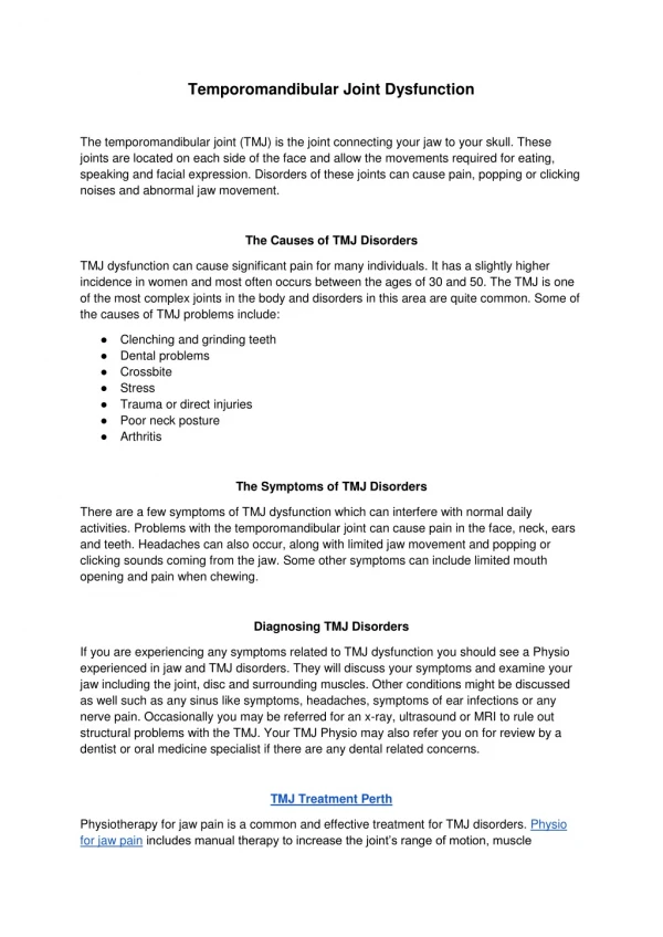
FUNCTIONAL ANATOMY OF TEMPOROMANDIBULAR JOINT. TEMPOROMANDIBULAR JOINT. Two components : PASIF COMPONENTS

E N D
TEMPOROMANDIBULAR JOINT Two components : PASIF COMPONENTS 1.Bone - Fossa mandibularis ossis temporalis - Capitulum mandibula ( condyle ) - Tuberculum articulare ( articular eminence )
2.Capsule and ligaments 3.Articular disc ACTIVE COMPONENTS Masticator muscles • Masseter • Temporalis • Pterygoideus medialis • Pterygoideus lateralis Additional muscles
Considered as a ginglymoarthrodial joint : - hinging movement ~ ginglymoid joint - gliding movement ~ arthrodial joint It is formed by mandibular condyle fitting into mandibular fossa and the two bones is separated by articular disc It is classified as a compound joint ( at least 3 bones ) ~ functionally the articular disc served as a nonossified bone
The TMJ is divided into superior and inferior cavity by the articular disc • Superior cavity - Superior : fossa mandibula Inferior : discus articularis - Gliding action between condyle and articular eminence • Inferior cavity - Superior : discus articularis Inferior : condyle - Hinge action between undersurface of the disc and the rotating surface of the condyle
MANDIBULAR CONDYLE = Processus condyloideus • It is the posterior portion of the ramus mandibula that extends upward • Mediolateral : 15 – 20 mm Anteroposterior : 8 – 10 mm • Anterior view : medial and lateral poles, the medial pole generally more prominent • The actual articulating surface ~ extends anteriorly and posteriorly to the most superior aspect ( P > A )
MANDIBULAR FOSSA • The squamous portion of the temporal bone ( concave ) • Anterior : a convex bony prominence ( tubercle ) = articular eminence • Posterior : squamotympanic fissure (M-L) ~ anteromedial : petrosquamous fissure ~ posteromedial : petrotympanic fissure
- The posterior roof is thin ~ not designed to sustain heavy force ~ in which condyle situated • The articular eminence consists of thick dense bone ~ to tolerate such forces - The steepness of the articular eminence surface ~ dictates the pathway of the condyle
CAPSULE AND LIGAMENTS 1.Capsula articularis ~ capsular ligament - It surrounds the entire TMJ - Superior attachment ~ the borders of the articular surface of the mandibular fossa and articular eminence Inferior attachment ~ collum mandibula - Function : ~ to resist any medial, lateral or inferior forces that tend to separate or dislocate the articular surface ~ to retain the synovial fluid
2.Collateral ( discal ) ligaments - From medial and lateral borders of the disc to the poles of the condyle ~ the medial discal ligament ~ the lateral discal ligament - Dividing the joint mediolaterally into superior and inferior joint cavities - Composed of collagenous connective tissue - Function : ~ allow the disc move passively with the condyle as it glides A - P
~ permit the disc to be rotated A-P on the articular surface of the condyle • These ligaments are responsible for the hinging movement between the condyle and the articular disc - They have a vascular supply and are innervated
3.Temporomandibular ligament - It lies at the lateral aspect of the capsular ligament - Composed of two parts : *Outer oblique portion From the outer surface of the articular tubercle and zygomatic process postero inferiorly to the outer surface of the condylar neck ~ it resists excessive dropping of the condyle so limiting the the extent of mouth opening
* Inner horizontal portion From the outer surface of the articular tubercle and zygomatic process posteriorly and horizontally to the lateral pole of the condyle and posterior part of the articular disc ~ it limits posterior movement of the condyle and disc
4.Sphenomandibular ligament - It is an accesory ligament - From the spine of the sphenoid bone and extends downward to lingula mandibula 5.Stylomandibular ligament - The second accesory ligament - From the styloid process and extends downward and forward to the angle and posterior border of the ramus mandibula - It limits excessive protrusive movements of the mandible
ARTICULAR DISC • Composed of dense fibrous connective tissue devoid of any blood vessels or nerve fibers • Sagittal plane ~ can be divided into 3 regions according to thickness - anterior border - posterior border ~ slightly thicker than anterior border - central area is the thinnest ~ intermediate zone ~ in which condyle is located normally
Anterior view ~ the disc is generally thicker medially than laterally ~ increased space between the condyle and the articular fossa toward the medial of the joint • The precise shape of the disc ~ morphology of the condyle and mandibular fossa - During movement the disc is somewhat flexible and can adapt to the functional demands of the articular surface ~ do not imply that morphology of the disc is reversibly altered during movement
The disc maintain its morphology unless destructive forces or structural changes occurs ~ its morphology can be irreversibly altered ~ biomechanical changes during function RETRODISCAL TISSUE • The articular disc is attached posteriorly to this region - It is a loose connective tissue region that highly vascularized and innervated
Superior : superior retrodiscal lamina ( contains many elastic fibers ) ~ bilaminary zone It attaches the disc posteriorly to the tympanic plate • Inferior : inferior retrodiscal lamina ( composed chiefly collagenous fibers ) It attaches the inferior border of the posterior edge of the disc to the posterior margin of the articular surface of the condyle - The remaining body of the tissue is attached posteriorly to a large venous plexus ~ it fills with blood as the condyle moves forward
Anterior region of the disc is attached to the capsular ligament - Superior : anterior margin of the articular surface of the temporal bone Inferior : anterior margin of the articular surface of the condyle - Composed of collagenous fibers Anteriorly the disc is also attached by tendinous fibers to the superior lateral pterygoid muscle
The articular surface of the mandibular fossa and condyle are lined with dense fibrous connective tissue ~ affords several advantages over hyaline cartilage : - less susceptible to the effects of aging ~ less likely to break down over time - a better ability to repair The internal surface of the joint cavity are surrounded by specialized endothelial cells that form a synovial lining ~ produces synovial fluid So TMJ ~ a synovial joint
The synovial fluid serves two purposes : • Acts as a medium for providing metabolic requirement, since the articular surfaces of the joint are nonvascular • As a lubricant during function Two mechanisms of the lubrication : • Boundary lubrication Prevents friction in the moving joint • Weeping lubrication Eliminates friction in the compressed but not moving joint
MUSCLES OF MASTICATION MASSETER • Rectangular muscle • There is two portions or heads ( caput ) Superficial head ( Caput superficial ) Origo : - processus zygomaticus ossis maxillae - 2/3 ventral of the inferior border of the zygomatic arch Insertio : extends downward and backward to the tuberositas masseterica
Profundus head ( Caput profundus ) Origo : - 1/3 dorsal of the inferior border of the zygomatic arch - medial surface of the zygomatic arch Insertio : extends downward and forward to ramus mandibula and lateral surface of processus coronoideus
As fibers of the masseter contract ~ mandible is elevated and the teeth are brought into contact • The superficial portion may also aid in protruding the mandible ~ the deep portion stabilize the condyle against articular eminence
TEMPORALIS • A fan-shaped muscle • Origo : - temporal fossa Its fibers extend downward between the zygomatic arch and the lateral surface of the skull Insertio : - processus coronoideus - ramus mandibula
Contraction of the muscle elevates the mandible and the teeth brought into contact • If only portions contract, the mandible is moved according to the direction of those fibers that are activated • According to fiber direction and ultimate function, it can be divided into 3 distinct areas :
* Anterior portion - The fibers are directed almost vertically - Contraction ~ mandible is raised vertically * Middle portion - The fibers run obliquely across the lateral aspect of the skull ( forward as they pass downward ) - Contraction ~ elevate and retrude the mandible
*Posterior portion -Run almost horizontally, coming forward above the ear to join other temporalis fibers as they pass under the zygomatic arch -The function is somewhat controversial ~ causes elevation and only slight retrusion
PTERYGOIDEUS MEDIALIS • Consists 2 heads ( caput ) Caput superficial Origo : - facies medialis lamina lateralis processus pterygoideus ( fossa pterygoideus ) - processus pyramidalis ossis palatina Caput profundus Origo : - processus pyramidalis ossis palatina - tuber maxillae
Extend downward, backward and outward to insert along the medial surface of the mandibular angle (tuberositas pterygoidea) - With the masseter, it forms a muscular sling ~ support the mandible - Contraction ~ mandible is elevated and the teeth are brought into contact - It is also active in protruding the mandible - Unilateral contraction ~ mediotrusive movement of the mandible
PTERYGOIDEUS LATERALIS It consists 2 heads or bellies with different function • Caput superior Origo : facies infratemporalis ala magna ossis sphenoidalis, extending almost horizontally, back ward and outward to insert on the : articular capsule, the disc and the neck of the condyle ( fovea pterygoid )
Caput inferior Origo : - facies lateralis lamina lateralis processus pterygoideus extends backward, upward and outward to insert on the neck of the condyle ( fovea pterygoidea )
Function : • The superior lateral pterygoid is active during power stroke ~ closure mandible against resistance ( chewing and clenching ) • While the inferior active during opening, the superior remains inactive, becoming active only in conjunction with the elevator • The right and left inferior contracts simultaneously ~ the condyles are pulled down the articular eminence and the mandible is protruded • The inferior functions with the mandibular depressors ~ the mandible is lowered and the condyles gide forward and downward on the articular eminences