Download
1 / 35
440 likes | 1.24k Vues
Radiographic examinations of the temporomandibular joint. Dobó-Nagy, Csaba Semmelweis University. Knowledge required by clinicians. Anatomy Available investigations Clinical indications How each investigation is performed position open or closed Limitations Pathological conditions.

E N D
Radiographic examinations of the temporomandibular joint Dobó-Nagy, Csaba Semmelweis University
Knowledge required by clinicians • Anatomy • Available investigations • Clinical indications • How each investigation is performed • position • open or closed • Limitations • Pathological conditions
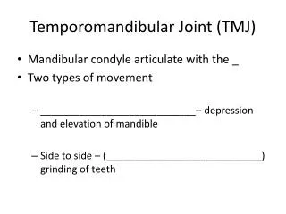
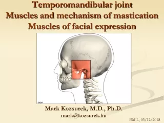
disk Glenoid fossa tubercle (articular eminence) condyle Anatomical section of a normal TMJ lateral pterygoid muscle
Skull base model A P R L
Main radiographyc projections of the TMJ TrCran TrOrb TrPhar RevTowne SMV
Limitations of routin radiographic projections • Soft tissue abnormalities • Effusion • Small and localised bony defects • One aspect of the joint (except for tomography)
A B C D E F G H I Main pathological signs of TMJ
Diagnostic informations of MR • Disk displacement • The integrity of the disk and its soft tissue attachments • Effusion (T2-weighted image) • Integrity of the articular surface • Research (gold standard)
Diagnostic informations of CT • The shape of the bony compartments • Information on all aspects of the joint • The condition of the articular surface • The size of the joint space • Integrity of the disk and soft tissue attachements
Suggested hierarchy of imaging modalities in TMJ disease soft tissue changes panoramic MRI bone changes tomography • Primary examination: • dysfunction • organic changes • failed treatment CBCT, CT