Download
1 / 36
490 likes | 930 Vues
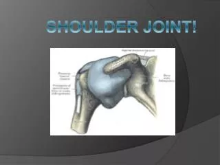
FUNCTIONAL ANATOMY OF SHOULDER JOINT. ARTICULATION. Articulation is between: The rounded head of the humerus and The shallow, pear-shaped glenoid cavity of the scapula. Head. Glenoid cavity. The articular surfaces are covered by hyaline cartilage.

E N D
ARTICULATION Articulation is between: • The rounded head of the humerus and • The shallow, pear-shaped glenoid cavity of the scapula. Head Glenoid cavity
The articular surfaces are covered by hyaline cartilage. • The glenoid cavity is deepened by the presence of a fibrocartilaginous rimcalled theglenoid labrum.
TYPE • Synovial • Ball-and-socket joint
FIBROUS CAPSULE • The fibrous capsule surrounds the joint and is attached: Medially to the margin of the glenoid cavity outside the labrum; Laterally to the anatomic neck of the humerus. • The capsule is thin and lax, allowing a wide range of movement.
LIGAMENTS Accessory ligaments: The coracoacromial ligamentextends between the coracoid process and the acromion. Its function is to protect thesuperior aspect of the joint. • Thecoracohumeral ligamentstrengthens the capsule from aboveand stretches from the root of the coracoid process to the greater tuberosity of the humerus. • Thetransverse humeral ligamentstrengthens the capsule and bridges the gap between the two humeral tuberosities. • The glenohumeral ligamentsare three weak bands of fibrous tissue that strengthen the front of the capsule.
SYNOVIAL MEMBRANE • It lines the fibrous capsule. • It isattached to the margins of the cartilage covering the articular surfaces. • It forms a tubular sheath around the tendon of the long head of the biceps brachii. • It extends through the anterior wall of the capsule to form the subscapularis bursabeneath the subscapularis muscle.
NERVE SUPPLY Articular branches of the axillary & the suprascapular nerves
The following movements are possible: • Flexion • Extension • Abduction • Adduction • Lateral rotation • Medial rotation Circumduction
Flexion • Normal flexion is about 90° • It is performed by the: • Anterior fibers of the deltoid • Pectoralis major • Biceps brachii • Coracobrachialis
Extension: • Normal extension is about 45° • It is performed by the: • Posterior fibers of the deltoid, • Latissimus dorsi • Teres major
Abduction: • Abduction of the upper limb occurs both at the shoulder joint and between the scapula and the thoracic wall. • It is initiated by supraspinatus from 0 to 18 • Then from 19 to 120 by the middle fibers of the deltoid. • Then above 90 by rotation of the scapula by 2 muscles ( Trapezius & S.A..)
The supraspinatus muscle: • initiates the movement of abduction(from 0 to 19) and • holds the head of the humerus against the glenoid fossa of the scapula; • This latter function of the supraspinatusallows the deltoid muscle to contract and abduct the humerus at the shoulder joint.
Adduction: • Normally the upper limb can be swung 45° across the front of the chest. • This is performed by: • pectoralis major • latissimus dorsi • teres major • teres minor
Lateral rotation: • Normal lateral rotation is about 40 to 45°. • This is performed by the: • infraspinatus • teres minor • the posterior fibers of the deltoid muscle
Medial rotation: • Normal medial rotation is about 55°. • This is performed by the: • subscapularis • latissimus dorsi • teres major • anterior fibers of the deltoid.
Circumduction: This is a movement in which the distal end of the humerus moves in circular motion while the proximal end remains stable • It is formed by flexion, abduction, extension and adduction. Successively
Posteriorly: • Infraspinatus • Teres minor muscles.
Superiorly: • Deltoid muscle • Coracoacromial ligament • Subacromial (subdeltoid) bursa • Supraspinatus muscle & tendon
the axillary nerve Inferiorly: • the posterior circumflex humeral vessels • the long head of the triceps muscle
The long head of the biceps brachii originates from the supraglenoid tubercle of the scapula, • It is intracapsular but extrasynovial • It's tendon passes through the shoulder joint and emerges beneath the transverse humeral ligament. • Inside the joint, the tendon is surrounded by a separate tubular sheath of the synovial capsule.
Abduction involves rotation of the scapula as well as movement at the shoulder joint. • For every 3° of abduction of the arm, a 2° abduction occurs in the shoulder joint and a 1° abduction occurs by rotation of the scapula. • At about 120° of abduction of the arm, the greater tuberosity of the humerus comes into contact with the acromion. • Further elevation of the arm above the head accomplished by rotating the scapula.
STABILITY OF THE SHOULDER JOINT • This joint is unstable because of the: • shallowness of the glenoid fossa • weak ligaments • Its strength almost entirely depends on the tone of the rotator cuff muscles. • The tendons of these muscles are fused to the underlying capsule of the shoulder joint. • The least supported part of the joint lies in the inferior location, where it is unprotected by muscles.
DISLOCATIONS OF THE SHOULDER JOINT The shoulder joint is the most commonly dislocated large joint. Anterior-Inferior Dislocation • Sudden violence applied to the humerus with the joint fully abducted pushes the humeral head downward onto the inferior weak part of the capsule, which tears, and the humeral head comes to lie inferior to the glenoid fossa.
A subglenoiddisplacement of the head of the humerus into the quadrangular space can cause damage to the axillary nerve. • This is indicated by paralysis of the deltoid muscle and loss of skin sensation over the lower half of the deltoid. • Downward displacement of the humerus can also stretch anddamage the radial nerve. Wrist drop
ROTATOR CUFF TENDINITIS • Lesions of the rotator cuff are a common cause of pain in the shoulder region. • Excessiveoverhead activity of the upper limb may be the cause of tendinitis, although many cases appear spontaneously. • During abduction of the shoulder joint, the supraspinatus tendon is exposed to friction against the acromion. • Under normal conditions the amount of friction is reduced to a minimum by the large subacromial bursa, which extends laterally beneath the deltoid.
Degenerative changes in the bursa are followed by degenerative changes in the underlying supraspinatus tendon, and these may extend into the other tendons of the rotator cuff. • Clinically, the condition is known as subacromial bursitis, supraspinatus tendinitis, or pericapsulitis. • It is characterized by the presence of a spasm of pain in the middle range of abductionwhen the diseased area impinges on the acromion.
RUPTURE OF THE SUPRASPINATUS TENDON In advanced cases of rotator cuff tendinitis, the necrotic supraspinatus tendon can become calcified or rupture.
Rupture of the tendon seriously interferes with the normal abduction movement of the shoulder joint. • The main function of the supraspinatus muscle is to hold the head of the humerus in the glenoid fossa at the commencement of abduction. • The patient with a ruptured supraspinatus tendon is unable to initiate abduction of the arm. • However, if the arm is passively assisted for the first 15° of abduction, the deltoid can then take over and complete the movement to a right angle.
SHOULDER PAIN • The synovial membrane, capsule, and ligaments of the shoulder joint are innervated by the axillary nerve and the suprascapular nerve. • The joint is sensitive to pain, pressure, excessive traction, and distension. • The muscles surrounding the joint undergo reflex spasm in response to pain originating in the joint, which in turn serves to immobilize the joint and thus reduce the pain. • Injury to the shoulder joint is followed by pain, limitation of movement, and muscle atrophy owing to disuse.
BRANCHES FROM THE SUBCLAVIAN ARTERY • The suprascapular artery, (branch from 1st part of subclavian artery) distributed to the supraspinous and infraspinous fossae of the scapula. • The superficial cervical artery, which gives off a deep branch that runs down the medial border of the scapula.
BRANCHES FROM THE AXILLARY ARTERY • The subscapular artery and its circumflex scapular branch supply the subscapular and infraspinous fossae of the scapula. • The anterior & posterior circumflex humeral artery. • Both the circumflex arteries form an anastomosing circle around the surgical neck of the humerus.
LIGATION OF THE AXILLARY ARTERY The existence of the anastomosis around the shoulder joint is vital to preserving the upper limb if it should it be necessary to ligate the axillary artery.