Download
1 / 59
600 likes | 763 Vues
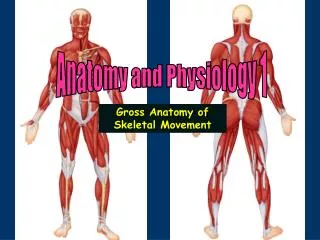
Anatomy and Physiology 1. Gross Anatomy of Skeletal Movement. The fleshy part of the muscle containing the muscle cells is the belly . At either end of the belly are the attachments, which are usually tendons that hold the muscle to the bones or other organs.

E N D
Anatomy and Physiology 1 Gross Anatomy of Skeletal Movement
The fleshy part of the muscle containing the muscle cells is the belly. • At either end of the belly are the attachments, which are usually tendons that hold the muscle to the bones or other organs. • Tendons (can be very long) allow the muscle to transmit force across a great distance. • Good example: wrists • Most of the muscles that move the fingers are found above the wrist. • These muscles are isolated by long tendons away from the point of movement. If all of our muscles were in our hands, our hands would be too large and movement would not be as effective nor could we handle things as delicately as we can.
Muscle contraction at the macroscopic level • Muscles are attached at each end to the skeletal system • Origin: the fixed (immovable) point of attachment. • Insertion: attachment to the movable bone • As a muscle shortens (contracts) theinsertionmoves toward theorigin
Muscle contraction at the macroscopic level • As a muscle shortens (contracts) the insertion moves toward the origin • Muscles can only pull they cannot push Direction of movement of radius towards scapula as biceps shortens
Antagonists oppose one another Pectoralis major “big chest muscle” Latissimus dorsi “big muscle on side of back” Pulls arm back and down. Used by swimmers Pulls arm forward and up
Look at distance between scapula and radius when the triceps are contracted The pink shows the additional length when the triceps are relaxed. Notice when the triceps are contracted that the elbow is no longer flexed and that the angle of the elbow joint is ~180 Triceps contracted
Look at distance between scapula and radius when the biceps are contracted The pink shows the additional length when the biceps are relaxed. Notice when the biceps are relaxed that the elbow is no longer flexed and that the angle of the elbow joint is ~180 Biceps contracted
Look at distance between tibia and femur when the quadraceps are contracted Insertion Quadraceps are knee extensors and will straighten the knee when they shorten
Look at distance between tibia and femur when the hamstrings are contracted Insertion Insertion Hamstrings are knee flexers and will bend the knee joint when they contract
Muscle contraction at the macroscopic level • Muscle can only pull they cannot push • Here the supraspinatus pulls on the greater tubercle of the humerus towards the posterior aspect of the scapula at the supraspinous fossa during abduction of the arm. It also helps to prevent the downward dislocation of the shoulder when carrying a heavy suitcase Relaxed Contracted
Approaches for learning the muscles. • Organize the muscles in your objectives by the joints that they move and learn them in groups. • Organize the muscles for each joint by the direction the move the joint (i.e., flex, extend, abduct) • Look at which bones and on what bone markings the muscles are attached. • Determine which is the origin and which is the insertion.
Bipedialism key to understanding limb movement Bipedialism (walking upright) believed to have first taken place between 3 - 5 million years ago provided for a division of labor between the limbs. • Upper Limb • Primarily structured for mobility and manipulation • Lost adaptation to support weight ( weight bearing) and/ or locomotion • Lower limb • Primarily structured for locomotion and weight bearing (support) • Slight ability to manipulate
Anatomy and Physiology 1 Movement of Arm/Shoulder Muscles
The upper limb is used for a broad range of both powerful and subtle actions, ranging from climbing, grasping, and throwing to writing, playing musical instruments and manipulating small objects. The major bones acted upon are: Scapula Humerus Radius Ulna Arm movements Abduction Adduction Flexion Extension Lateral rotation Medial rotation Elevation Depression Retraction Protraction Pronation Supination Movements of the upper limb
Coracovicular joint Acromioclavicular joint clavical Glenohumeral joint Sternoclavicular joint scapula humerus Scapulothoracic joint Arm muscles move against the joints of the pectoral girdle (shoulder), elbow and wrist
Posterior muscles that act on the pectoral girdle Levator scapulae Trapezius Rhomboid minor Rhomboid major
Anterior muscles that act on the pectoral girdle Levator scapulae Trapezius Rhomboid minor Rhomboid major
Anterior muscles that act on the pectoral girdle Pectoralis minor Serratus anterior
Deltoid Coracobrachialis Pectoralis minor Teres major 3 scapular 2 axial Latissimus dorsi Nine muscles act on humerus 4 rotator cuff Teres minor Supraspinitus Infraspinitus Sucapsularis
Axial muscles acting on the humerus attach arm to trunk and are prime movers of shoulder
Rotator cuff muscles reinforce the joint capsule and hold humerus in the glenoid cavity Supraspinatus Infraspinitus Teres minor
Three rotator cuffs muscles pull the greater tubercle of the humerus toward the scapula Supraspinitus assists in abduction of the arm. Relaxed Contracted Infraspinitus rotate humerus laterally and prevents humerus from sliding upwards Teres minor rotates humerus laterally, prevents humerus from sliding upwards and modulates action of deltoid
Remaining shoulder muscles are scapular Deltoid Coracobrachialis Teres major
Muscle of the shoulder: pectoralis major When the pectoralis major shortens insertion moves toward origin and the arm is pulled across the chest is a movement that adducts the arm and flexes the shoulder Insertion: intertubercular groove of humerus Origins: 1. sternal end of clavicle 2. sternum, cartilage of ribs 1-6 or 7, 3. apponeurosis of external oblique muscle
Deltoid (large, fleshy, triangular muscle that covers the shoulder and causes the bulge of the upper arm) runs from both the clavicle and the scapula to the humerus. Origin Insertion When the deltoid shortens insertion moves toward origin Arm is pulled away from body (i.e., abducted) Arm is horizontal when the deltoid is contracted fully
Shoulder movement: latissimus dorsi Latissimus dorsi is a wide, triangular muscle of the lower back. This muscle originates from the lower spine and sweeps upward to insert on the humerus. to extend and adduct the arm. This muscle is very important for swimming, rowing, and climbing a rope. Insertion: spirals around teres major to insert into the intertubercular groove of humerus Origin: indirect attachment via lumbodorsal fascia into spines of lower 6 thoracic vert., lumbar vert.,lower 4 to 4 ribs and iliac crest
Muscles of the shoulder/elbow: biceps brachii Origin Biceps brachii is a muscle of the anterior upper arm that is familiar because it bulges when the forearm is flexed. It also supinates the hand when a doorknob is turned or a cap of a jar is unscrewed. The name of the muscle refers to its two heads that attach to the scapula, where it originates. The biceps brachii inserts on the radius. Insertion
Muscles of the shoulder/elbow: triceps brachii Origins: scapula (long head); posterior shaft of humerus (lateral head); posterior humeral shaft distal to radial groove (medial head) Triceps brachii is the only muscle of the posterior upper arm. It has three heads that attach to the scapula and humerus, and it inserts on the ulna. The triceps extends the forearm when something is pushed. It is sometimes called the boxer's muscle because it straightens the elbow when a punch is thrown. Insertion: by common tendon into olecranon process of ulna
Muscles of the shoulder/elbow: triceps brachii Contraction of triceps straightens arm and pulls the elbow open
Muscles of the wrist: flexor carpi radialis Flexor carpi radialis muscle originates on the bones of the forearm and insert on the bones of the hand. They move the wrist and hand. Notice this muscle is on the anterior side of the arm Origin: medial epicondyle of humerus Insertion: base of second and third metacarpals
Muscles of the wrist: flexor carpi radialis Origin Insertion When the flexor carpi radialis shortens the insertion moves toward origin and the wrist flexes The hand moves out of the plane of the image towards you-bending the wrist downwards to the forearm
Muscles of the wrist: extensor digitorum The extensor digitorum muscle originates on the bones of the forearm and insert on the bones of the hand. They move the fingers. Notice that this muscle is on the posterior side of the arm Origin: Lateral epicondyle of humerus Insertion: medial 4 digits
Muscles of the wrist: extensor digitorum Origin Insertion When the extensor digitorum shortens the insertion moves toward origin and the wrist flexes The hand moves out of the plane of the image towards you bending the wrist backwards
Anatomy and Physiology 1 Movement of Hip/Thigh/Leg Muscles
Muscles that Move the Thigh Muscles of the thigh and lower limb tend to be large and heavy because they are used to move the entire weight of the body and resist the force of gravity. Therefore, they are important for movement and balance.
Muscles that Move the Thigh Iliopsoas originates from the ilium and the bodies of the lumbar vertebrae, and inserts on the femur anteriorly. This muscle flexes the thigh and is important to the process of walking. It also helps prevent the trunk from falling backward when a person is standing erect.
Muscles that Move the Thigh Origin: anterior superior iliac spine The Sartorius is a long, straplike muscle that begins at the iliac spine and then passes inward across the front of the thigh to descend over the medial side of the knee. It flexes the leg and is used to sit cross-legged, as tailors were accustomed to do in another era. Therefore, it is sometimes called the tailor's muscle, and, in fact, sartor means tailor in Latin. Insertion: winds around medial aspect of knee and inserts into medial aspect of proximal tibia
Sartiorus Because the origin is above the hip as the muscle shortens the hip will bend (flexion) Because the muscle crosses from the lateral to the medial side of the leg, when the muscle shortens the leg will twist outward (laterally rotate) as insertion moves to origin. Because the insertion is below the knee as the muscle shortens the knee will bend (flexion)
Arrow indicate direction of femur movement when adductors contract Muscles that Move the Thigh Adductor muscles are located on the inner thigh (medial aspect). They originate from the pubic bone and ischium, and insert on the femur. Adductor muscles adduct the thigh and press the thighs together. As the muscles shorten they will pull the medial femur toward the midline
Note: this is the back of the leg Muscle that Move the Thigh Origin: inferior ramus and body of pubis The Gracillis muscles are located on the medial parts of each thigh. They originate from the pubic bone and ischium, and insert on the femur. Adductor muscles adduct the thigh and press the thighs together. The Gracilis assists the Sartorius in flexing the leg and rotating it inward; it is also an adductor of the thigh. If the lower extremities are fixed, these muscles maintain the body in an erect posture and if contracted can flex the pelvis forward upon the femur Insertion: anterior medial tibial condyle
Muscles that Move the Lower Limb The quadraceps is a powerful knee extensor used in climbing, jumping, running, and rising from a seated position. This muscle group arises from 4 heads that form the flesh of the front sides of the thigh (rectus femoris, vastus lateralis, vastus medialis, vastus intermedius)
Origin: anterior inferior iliac spine and superior margin of acetabulum Insertion: patella and tibial tuberosity via patellar ligament Muscles that Move the Thigh Quadriceps femoris group includes muscles found in the front and on the sides of the thigh. The rectus femoris, which originates from the ilium, lies in front of the vastus intermedius. The vastus muscles originate from the femur. They are primary extensors of the lower leg, such as when a ball is kicked.