Download
1 / 14
300 likes | 1.39k Vues
The Liver: Blood supply, Portal Hypertension and Portosystemic Anastomoses. Rachael Edgar and Ellie Borton. The blood supply of the Liver. The liver receives blood from two sources. What are they? Arterial supply from Hepatic Artery (30%) Venous supply from Hepatic Portal Vein (70%)

E N D
The Liver:Blood supply, Portal Hypertension and Portosystemic Anastomoses Rachael Edgar and Ellie Borton
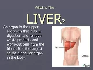
The blood supply of the Liver • The liver receives blood from two sources. What are they? • Arterial supply from Hepatic Artery (30%) • Venous supply from Hepatic Portal Vein (70%) • The Hepatic Portal vein comes from? • Splenic vein • Superior mesenteric vein
What are the components of the portal triad? • Bile duct • Portal vein • Hepatic artery The portal vein and hepatic artery drain into the central vein.
Acini and Lobules • Hepatic Acinus • Functional unit liver • Important is Line connecting 2 portal triads & extends out to 2 adjacent central veins • Dif areas oxygenation • Hepatic Lobule • Small physical division of the liver • Hexagon with vein in the centre, periphery has portal triads
Where does the central vein drain to? • Hepatic vein
Portal Hypertension • What happens? • Prolonged damage to the hepatocytes causes fibrosis (scarring) • Blood supply via the hepatic portal vein is blocked and the pressure within it rises • This causes a ‘back up’ of blood in the hepatic portal vein • Collaterals occur within the systemic venous system
What are the main sites of the collaterals? • Azygous veins (at gastro-oesophageal junction) • Rectum • Anterior abdominal wall via umbilical vein
What are the clinical consequences of portal hypertension? • Oesophageal varices • Rectal varices • Caput medusa
Rectal Varices • These are found higher in the rectal canal (found in mid rectum) than haemorrhoids.
Haemorrhoids • Common condition caused by ‘disrupted & dilated’ anal cushions - symptomatic • May be classified as internal or external in relation to pectinate line • Symptoms of bleeding, pain or itch, may be due to enlargement, inflammation, prolapse orthrombosis