Download
1 / 40
490 likes | 1.64k Vues
Complications in refractive surgery. Modified by Corina van de Pol, O.D., Ph.D. July 28, 2001 James Colgain, OD Mitch Brown, OD, FAAO. Complications of PRK. refractive over/undercorrection Regression Central Islands laser Decentered ablation. healing Epithelial compromise

E N D
Complications in refractive surgery Modified by Corina van de Pol, O.D., Ph.D. July 28, 2001 James Colgain, OD Mitch Brown, OD, FAAO
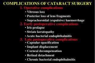
Complications of PRK • refractive • over/undercorrection • Regression • Central Islands • laser • Decentered ablation • healing • Epithelial compromise • Corneal infection • Corneal haze • Corneal scar • infection - rare 1:5,000
Over/under-correction • Cause: • inaccurate refraction • unstable • CTL warpage especially in HCL or GPHCL • undetected pathology (KCN) • unpredictable healing • induced cylinder • occult autoimmune disorder
Over/under-correction • Treatment: • based on refractive stability • change no greater than 0.5D over 1 month • wait longer in higher myopes and hyperopes • most surgeons wait at least 3 months • ok to treat interim over-correction with SCL • UCVA >20/40 • based on expectations, patient desire
Regression • <0.25D myopic regression over 1 year • US Navy study (n=100) • Retreatment possible • based on refractive stability and visual symptoms/complaints
Central islands • Cause: • plume/debris • water • Rare in Lasik, rarer still with scanning lasers • Not as prevalent with newer software and scanning lasers
Central islands • Treatment: • observe • >90% of islands resolve spontaneously • customized ablation • based on height and diameter of island
Epithelial compromise • Cause: • underlying basement dystrophy • prior trauma • dry eye • smoking
Epithelial compromise • Treatment: • patient selection • copious tears • consider punctal occlusion • bandage CTL • proper fit • Acuvue 8.8 for K<40 • Acuvue 8.4 for K>40 • topical antibiotic until epithelium healed
Corneal infection • Rare (<1:5,000) • Worked up and treated like CTL-related microbial keratitis • if <2 mm, mid-periphery to limbus, consider empiric therapy with fluoroquinolone • if >2 mm and/or central/paracentral, consider scraping for culture and sensitivity and aggressive topical fortified antibiotics (cefazolin and tobramycin)
Corneal haze • Cause: • unclear • ? UV exposure • ? Over-exuberant healing response
Corneal haze • Treatment: • unclear • based on vision and refraction • probably no treatment required if not visually significant • if K's are steepening and refraction shifting toward myopia, consider trial of FML • >95% of haze clears eventually
Corneal scar • Unresolved haze, refractory to FML • Potential for vision loss • Consider PF • Consider corneal scraping • Consider mitomycin-C or thio-tepa
refractive over/undercorrection induced astigmatism central islands - rare laser decentered less with tracking more with longer ablations angle kappa and visual axis? flap buttonhole in pupil free cap if small epithelial defects especially with older patients and dry eyes wrinkles, striae decentration inflammation (DLK) epithelial ingrowth (primary and secondary) infection - rare 1:5,000 Complications of LASIK
Buttonhole • Cause: • steep K (>46), greater risk • cornea "buckles" during microkeratome pass, creating central area where blade exits cornea then re-enters. This is often in the visual axis and is disastrous to vision if the ablation occurs. • May re-cut deeper in cornea in 3-6 months
Buttonhole • Treatment: • do NOT perform laser ablation • irregular astigmatism WILL be induced • replace flap or don’t lift at all • allow cornea to heal (at least 3 months) • re-cut thicker flap and decenter entry of the MK so as not to disturb initial plane
Free cap • Cause: • flat K (<40D) these are at greater risk • microkeratome travels completely across flap • no hinge created • ALK used to be performed in this fashion • Surgeon MAY proceed if he bed, cap and area for ablation are normal • Always necessary to mark cornea so the epi side is placed up when repositioned
Free cap • Treatment: • save free cap in antidessication chamber • complete laser ablation • replace cap, aligning with preplaced marks, epithelium UP • consider suture (usually not required) and bandage CTL
Epithelial defects • Cause: • pre-existing condition • ABM dystrophy • recurrent erosion • prior trauma • dry eye • greater suction and torquing motion • dry surface during microkeratome pass
Epithelial defects • Treatment: • patient selection • pre-existing epithelial conditions listed above are relative contraindications to LASIK. • Consider surface PRK for above conditions • copious irrigation during procedure • wet cornea just prior to keratome pass • bandage CTL • intraoperative defects may end up being areas of RCE during healing phase
Flap striae • May result in irregular astigmatism and lost BCVA • Cause: • technique • flap laid back with poor attention to detail • not smoothed properly • more significant in higher myopes • patient • rubbed eye/flap during day one to week one • possible to dehisce flap completely in first 24-48 hours
Flap striae • Treatment: • technique • meticulous attention to smoothing flap at time of ablation and positioning with attention to “gutter” and pre-op marker alignment • consider "pressing" flap • consider refloating flap if visually significant • rarely, suturing required to stretch flap • patient • clear shield at night for first week • caution patient not to rub eye • use tears for irritation
Flap de-centration • Cause: • eye torques when suction applied • may result in decentered ablation • the larger the ablation zone - especially in hyperopes the more significant this issue • prior to treatment, the surgeon may view the area of ablation on most lasers to determine whether the bed area is OK for the treatment
Flap decentration • Treatment: • if ablation can be performed without hitting flap edge, consider proceeding • if ablation cannot be accomplished without hitting flap edge, abort laser, replace flap, allow cornea to heal (at least 3 months) and recut deeper, centered flap possibly using a different MK
Inflammation • Received the most press as potential complication following LASIK • Called many names: • Diffuse lamellar keratitis (DLK) • Sands of the Sahara syndrome (SOS) • May occur in “groups or outbreaks” • Causes (many potential, none proven): • metallic debris, meibomian secretions, staph toxin, keratome oil, infection
Inflammation • Treatment (stage-dependent): • stage I: increased frequency of FML • stage II: switch to Pred Forte • stage III: lift flap, irrigate and add Pred Forte • stage IV: stage III Rx and pray • vision loss probable
Epithelial ingrowth • Causes (2 types): • nests of cells deposited under flap during procedure • migration of epithelium at flap edge
Epithelial ingrowth • Treatment: • observe for progression • if progressive, lift flap, scrape with Weckcell, irrigate well and reposition flap • may require lifting flap more than once • risk of epithelial ingrowth increases each time flap is lifted • More risk with older patients and poorer epithelium • follow up, early detection and treatment critical to the best outcome
Infection (lamellar keratitis) • Potentially the most devastating complication associated with LASIK • Fortunately, a rare complication (<1:5,000) • Causes: • poor Betadine prep • poor lid/lash drape • bad luck • post op introduction of infectious agent
Infection • Treatment: • consider lifting flap to scrape for culture and sensitivity • consider aggressive topical fortified antibiotics (cefazolin and tobramycin)
Flap Dislodgment after Lasik • Rare: no real studies just reported events • No one knows when the flap heals • Able to lift some patients 3 years out • Events leading to flap dislodgment or striae from trauma after 30 days • Airbag, cat and dog scratch, cardboard box edge, fingernail scratch during fight, retinal buckle surgery, tree branch hitting cornea, snowball hit eye
PRK • Advantages • safer • longer track record • costs less • Disadvantages • slower recovery • more discomfort • corneal haze • limited range
LASIK • Advantages • faster recovery • less discomfort • less follow-up • enhancements easier • high myopia • Disadvantages • increased risk • late flap displacement • increased cost
PRK vs. LASIKSame destination; Different journey • PRK • Day one: “Oowww!” • Less surgical risk • Slower recovery • 80% 20/20 • Haze • No flap • 0.2 – 0.3% risk visual loss (>2 lines) • LASIK • Day one: “Wow!” • Greater surgical risk • Quicker recovery • 80% 20/20 • No haze • Flap • 0.2 – 0.3% risk visual loss (>2 lines)