Download
1 / 31
560 likes | 2.73k Vues
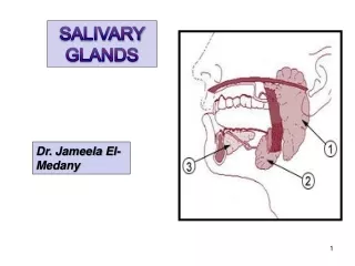
Salivary Glands Disorders. Anatomical Considerations. Two submandibular Two Parotid Two sublingual > 400 minor salivary glands. Minor salivary glands. These lie just under mucosa. Distributed over lips, cheeks, palate, floor of mouth & retro-molar area.

E N D
Anatomical Considerations • Two submandibular • Two Parotid • Two sublingual • > 400 minor salivary glands
Minor salivary glands • These lie just under mucosa. • Distributed over lips, cheeks, palate, floor of mouth & retro-molar area. • Also appear in upper aerodigestive tract • Contribute 10% of total salivary volume.
Sublingual Salivary glands • This is the smallest of the major salivary glands. • The almond shaped gland lies just deep to the floor of mouth mucosa between the mandible & Genioglossus muscle. • It is bounded inferiorly by the Mylohyoid muscle • Sublingual gland has no true fascial capsule. • It lacks a single dominant duct. Instead, it is drained by approximately 10 small ducts (the Ducts of Rivinus)
Submandibular Gland • This gland lies in the submandibular triangle formed by the anterior and posterior bellies of the Digastric muscle and the inferior margin of the mandible. • The gland forms a ‘C’ around the anterior margin of the Mylohyoid muscle, which divides the gland into a superficial and deep lobe.
Submandibular Gland…… • Wharton’s duct empties into the intraoral cavity lateral to the lingual frenulum on the anterior floor of mouth
Parotid Gland • The parotid gland represents the largest salivary gland • The following lists the boundaries of the parotid compartment: •Superior border – Zygoma •Posterior border – External Auditory Canal •Inferior border – Styloid Process, Styloid Process musculature, Internal Carotid Artery, Jugular Veins •Anterior border – a diagonal line drawn from the Zygomatic root to the EAC
Parotid Gland…… • 80% of the gland overlies the Masseter and mandible. The remaining 20% of the gland (the retromandibular portion • This portion of the gland lies in the Prestyloid Compartment of the Parapharyngeal space
Parotid Gland…… • Stensen’s duct arises from the anterior border of the Parotid and parallels the Zygomatic arch, 1.5 cm inferior to the inferior margin of the arch. • It runs superficial to the masseter muscle, then turns medially 90 degrees to pierce the Buccinator muscle at the level of the second maxillary molar where it opens onto the oral cavity.
Parotid Gland…… • Cranial Nerve VII divides it into 2 surgical zones (the superficial and deep lobes). • After exiting the foramen, it turns laterally to enter the gland at its posterior margin. • The nerve then branches at the Pes Anserinus (goose’s foot) approximately 1.3 cm from the stylomastoid foramen. The nerve then gives rise to 2 divisions: • 1)Temperofacial (upper) • 2)Cervicofacial (lower)
Parotid Gland…… • Followed by 5 terminal branches: • 1)Temporal • 2)Zygomatic • 3)Buccal • 4)Marginal Mandibular • 5)Cervical
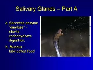
Functions of saliva include the following: • It has a cleansing action on the teeth • It moistens and lubricates food during mastication and swallowing • It dissolves certain molecules so that food can be tasted • It begins the chemical digestion of starches through the action of amylase, which breaks down polysaccharides into disaccharides. • The saliva from the parotid gland is a rather thin, watery fluid, but the saliva from the sublingual and the submandibular glands contains mucus and is much thicker.
Disorders of minor salivary Glands • Extravasation Cysts • Follow trauma • MSG with in lower lip • Visible painful swelling • Some resolve spontaneously or require surgery
Disorders of minor salivary Glands • MSG tumours are rare but 90% are malignant • Common sites include • Upper lip • Palate • Retromolar regions • Rare sites are nose/PNS/Pharynx
Disorders of minor salivary Glands • Benign tumours present as painless slow growing swellings, overlying ulceration is rare. • Malignant tumours have firmer consistency and have ulceration at later stage
Disorders of minor salivary Glands • Benign tumors of palate < 1cm in size are removed by excisional biopsy • When size larger than 1 cm prior incisional biopsy is done • Malignant tumors are managed by excision which may involve low-level or total maxillectomy and immediate reconstruction
Disorders of sublingual salivary Glands • Problems are rare • Minor mucous retention cysts • Plunging ranula is a retention cyst that tunnels deep • Nearly all tumours are malignant
Plunging ranula • Rare form of retention cyst • May arise from SM/SL SG • Mucous collects around gland • Penetrates Mylohyoid muscle to enter neck • Soft painless fluctuant dumb-bell shaped swelling • Surgical excision via neck
Disorders of sublingual salivary Glands • Tumours are rare • 90% are malignant • Wide excision and simultaneous neck dissection
Acute sialadenitis Viral (Mumps) Bacterial secondary to infection More Common Secondary to obstruction Poor capacity to recover Despite control with Abx chronicity follows and requires surgical excision Disorders of submandibular salivary Glands
Chronic Sialadenitis • Commonly due to obstruction following stone formation • 80% salivary stones occur in SMSG • High mucous content • Acute painful swelling rapidly precipitated by eating & resolves within 1-2 hours • Enlarged bimanually palpable SMG • Marsuplisation/Excision
Tumors of Submandibular Salivary Glands • Uncommon, slow growing, painless • Only 50% are benign • Even malignant tumours can be slow growing • Pain is not a reliable feature • Investigations: • CT/MRI • FNAC • No open biopsy
Management • Small & encased within capsule intracapsular excision • Large benign tumors– suprahyoid excision • Malignant tumours require concomitant neck dissection
Disorders of parotid Glands • Common causes of parotid swelling: • Mumps • Acute bacterial sialadenitis in dehydrated elderly patients • Acute bacterial parotitis • Obstructive parotitis: causes swelling at meal time
Parotid Tumours • Most Common is pleomorphic adenoma (80-90%) • Low grade Tumors like acinic cell carcinoma are not distinguishable from benign • High grade Tumours grow rapidly, are often painful and have nodal metastasis • CT/MRI are useful • FNAC better than open biopsy • Tx should be excised & not enucleated
Classification of Parotid Tumours • Adenoma • Pleomorphic • Monomorphic (Warthin’s Tumour) • Carcinoma • Low grade (Acinic cell/Adenoid cystic) • High grade (Adenocarcinoma/SCC)
Management • Superficial parotidectomy most common procedure • Radical parotidectomy is performed for patients clear histological evidence of high grade malignancy
Tumour like lesions • Sialadenosis • Diabetes • Alcoholism • Endocrine disorders • Pregnancy • Bulimia
Sjogren Syndrome • Autoimmune condition causing progressive degeneration of salivary and lachrymal glands • The oral aspects of primary Sjogren's syndrome consist of mucosal atrophy (80% to 95%), salivary gland enlargement approximately 30 %), • The oral manifestations may include xerostomia with or without salivary gland enlargement, candidiasis, dental caries and taste dysfunction.
Investigations • Sialometry • Sialography • Scintigraphy a radioactive tracer is given by vein that is subsequently taken up by the salivary glands and gradually eliminated within the salivary fluid • Sialochemistry • Ultrasonogram • Labial or minor salivary gland biopsy
Management • Symptomatic • From the systemic drug treatment standpoint, immunosuppressive therapy in the form of corticosteroids or cytotoxic drugs have proven effective, in particular when symptoms are severe. A drug known as Plaquenil has also proven to be helpful in some cases with open questions remaining as to the role of alpha interferon and nonsteroidal anti-inflammatory drugs.