SALIVARY GLANDS
240 likes | 6.21k Vues
SALIVARY GLANDS. Objectives By the end of the lecture the student should be able to: Describe the microscopic structure of the major salivary glands in correlation with function. SALIVARY GLANDS. (A) Major Salivary Glands : 1- Parotid. 2- Submandibular. 3- Sublingual.

SALIVARY GLANDS
E N D
Presentation Transcript
SALIVARY GLANDS Objectives By the end of the lecture the student should be able to: • Describe the microscopic structure of the major salivary glands in correlation with function.
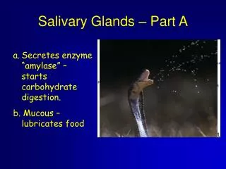
SALIVARY GLANDS (A) Major Salivary Glands: 1- Parotid. 2- Submandibular. 3- Sublingual. (B) Minor Salivary Glands: • Labial, Lingual, Buccal, Palatine. • Produce 5% of salivary output.
Major Salivary Glands • Stroma: • C.T. capsule. • C.T. septa dividing the glands into lobes and lobules. • Parenchyma: • Acini. • Duct system.
Types of Salivary Acini • Serous Acini: • Contain only serous cells. • Small, spherical, and with a narrow lumen. • Secrete serous secretion rich in enzymes, such as amylase and lysozyme. • Mucous Acini: • Contain only mucous cells. • Larger, more tubular, and with a wider lumen. • Secrete mucous secretion. • Mucoserous (Mixed) Acini: • Mucous acini with a cap of serous cells (serous demilunes).
Types of Salivary Acini Mucous Serous Mixed
Cells of Salivary Acini 1. Serous cells 2. Mucous cells Pyramidal or cuboidal. Nuclei are flattenedand basal. Cytoplasm: Pale basophilic and vacuolated (foamy) (due to dissolved mucinogen secretory granules). • Pyramidal in shape. • Nuclei are roundand basal. • Cytoplasm: • Deeply basophilic (due to numerous RER), with apical acidophilic secretory granules (rich in salivary amylase). 3. Myoepithelial cells (basket cells): • Contractile cells that embrace the basal aspect of the acini. • Their contraction releases the secretion into the duct system.
Duct System of Salivary Glands • Intralobular ducts (prominent): • Intercalated ducts: • lined by small cuboidal cells. • Striated ducts: • lined by low columnar cells. • Interlobular ducts: • lined by simple columnar epithelium. • Main duct: • lined by stratified columnar epithelium which becomes stratified squamous (nonkeratinized) in the distal end.
Parotid Gland • The largestsalivary gland. • Produces 30% of salivary output. • Purely serous. • Prominent intralobular ducts. • Secretion rich in: • Amylase. • Lactoferrin. • Lysozyme. • Secretory IgA.
Submandibular Gland • Produces 60% of salivary output. • Mixed but mostly serous (90%). • Mucous acini are capped by serous demilunes.
Sublingual Gland • The smallest salivary gland. • Produces 5% of salivary output. • Mixed but mostly mucous. • Mucous acini are capped by serous demilunes.
Parotid: purely serous Submanddibular: mostly serous Sublingual: mostly mucous