Download
1 / 15
150 likes | 247 Vues
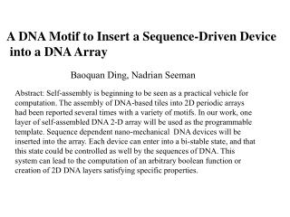
Explore e-motif structure in DNA, focusing on extra-helical cytosines and their role in Fragile X syndrome. Learn about project objectives and methods used to model and analyze these structures. Acknowledgments to project contributors.

E N D
eE-Motif Structures in DNA Kathryn Iverson USC Health Sciences Center and School of Pharmacy SoCalBSI 2007
Outline • E-motif structure • Project objectives • Methods • Working results
G C C G C C 5’ CGCCGCCG CCGCCGCC G C CGCCGCCG CCGCCGCCG C G GGCGGCGGC C GGCG G GCGGCGGCGGCGGCGG CGGCGGC 5’ C G C C G G C G G C G G G C Hairpins formed by GCC repeats
5’ C GC CG GC CG C 5’ C C
E-motif Structure • Extra-helical cytosines • Extra-helical cytosines from first GCC and third GCC repeats are interacting • Stacking between G-C
Fragile X • Is the most common forms of inherited mental retardation. 1 in 2000 boys and 1 in 4000 girls are afflicted and 1 in 260 women are carriers1 • July 22, National Fragile X Awareness Day • Characterized by the expansion of GCC repeat in the 5’ untranslated region of the FMR1 gene 1. http://www.conquerfragilex.org/about.php
Objectives of Project • Accurate model of eE-motif structure • Knowledge of extra-helical cytosine structures • Insight into Fragile X syndrome • Therapeutic targets
Methods • Generate NASDAC input files • Run energy minimization on the structures using AMBER • Molecular dynamics modeling with restraints in AMBER
Generation of pdb files • User enters how many GCC repeats • Option to protonate one of cytosines • What parameter to vary and within what range • Create data.in file, run NASDAC and produce .pdb file
Minimization Without Restraints • G-C base pairs no longer close enough to hydrogen bond • Stacking of extra-helical cytosines
Restraints • Hydrogen bonding distance between G and C base pairs • Distance between N2 atoms in G-C base pairs • Torsion angle for G-C base pairs • Distance between extra-helical cytosines
eE-Motif with Restraints • Extra-helical cytosines are in the same plane • Hydrogen bonding
Acknowledgements • Thank you to Dr. Ian Haworth and Dr. Rebecca Romero for their guidance and advice throughout the project • USC Health Sciences Center and School of Pharmacy • Program leaders and faculty of SoCalBSI • SoCalBSI participants • Cal State LA