Cardiac Lecture
Cardiac Lecture. Jan Bazner-Chandler CPNP, CNS, MSN, RN. Cardiac. Ball & Bindler. Focused Health History. Family history of defects / early cardiac disease / siblings with defects Maternal history of stillborns or miscarriages

Cardiac Lecture
E N D
Presentation Transcript
Cardiac Lecture Jan Bazner-Chandler CPNP, CNS, MSN, RN
Cardiac Ball & Bindler
Focused Health History • Family history of defects / early cardiac disease / siblings with defects • Maternal history of stillborns or miscarriages • Congenital anomalies / genetic anomalies / fetal alcohol syndrome / Down Syndrome and Turner Syndrome • Maternal exposure to rubella
Focused Health History • Heart murmur • Tires while eating • Low weight for height • Sweats while eating (diaphoretic) • Cyanosis, worsens with feeding or activity level • Irritable weak cry
Focused Health History • In the older child additional symptoms may include: • Chest pain • Decreased activity level • Syncope • Slight of build
Focused Physical Assessment • General appearance • Integumentary system • Face, nose, and oral cavity • Thorax and lung • Cardiovascular system
Heart Murmurs • These sounds are produced by blood passing through a defective valve, great vessel, or other heart structure. • Murmurs are classified by: intensity, location, radiation, timing, and quality.
Pulses • Alert: • Weaker pulses or lower blood pressure in the lower extremities may indicate coarctation of the aorta (COA) • Bounding pulses can indicate a patent ductus arteriosus (PDA) or aortic insufficiency.
Vital Signs • Heart rate: tachycardia in the absence of fever, crying, or stress may indicate cardiac pathology. • Tachypnea, even with rest, chest retractions indicate respiratory distress, possibly resulting from congestive heart failure
Knee-chest Position Nurse puts infant in knee-chest position. Whaley & Wong Child with a cyanotic heart defect squats (assumes a knee-chest position) to relieve cyanotic spells. Some times called “tet” spells. Ball & Bindler
First Breath • Pulmonary alveoli open up • Pressure in pulmonary tissues decreases • Blood from the right heart rushes to fill the alveolar capillaries • Pressure in right side of heart decreases • Pressure in left side of heart increases • Pressure increases in aorta
Treatment Modalities • Palliative procedures • Pulmonary artery banding • Shunts • Corrective procedures
Diagnostic Test • Chest x-ray to define silhouette of the heart. • Heart size, shape, pulmonary markings, and cardiomegaly. • Electrocardiogram ECG or EKG to define electrical activity of the heart. • Echo-cardiogram to visualize anatomic structures. Non-invasive
Cardiac Catheterization • An invasive test to diagnose or treat cardiac defects. • Visualizes heart and vessels. • Measures oxygen saturation of chambers. • Measures intra-cardiac pressures. • Determines muscle function and pumping action of the heart.
Toxicity to Dye • Watch for signs of toxicity to the dye used during the procedure. • Increased temperature • Urticaria • Wheezing • Edema • Dyspnea • Headache *Allergy response
Pre-cardiac Catheterization • Assess vital signs with blood pressure. • Hemoglobin and hematocrit • Pedal pulses • NPO • Hold digoxin • IV if child is polycythemic
Post-cardiac Catheterization • Vital sign, with apical pulse, and blood pressure q 15 minutes for first hour. • Apical pulse for 1 minute to check for bradycardia or dysrhythmias.
Post-cardiac Catheterization • Assess pulses below the cath site. • Record quality and symmetry of pulses. • Assess temperature and color of affected extremity. • Check dressing for bleeding or hematoma formation.
Home Care Instructions • Keep dressing in place for 24 hours. • Keep site dry and clean. • Observe site for redness, swelling, drainage, or bleeding. • Check temperature. • Avoid strenuous exercise. • Acetaminophen for pain. • Keep follow-up appointment • Pre-procedure medications as ordered.
Left to Right Shunt • Pressures on the left side of the heart are normally higher than the pressures in the right side of the heart. If there is an abnormal opening in the septum between the right and left sides, blood flows from left to the right.
Clinical Manifestations • The infant is not cyanotic. • Tachycardia due to pushing increased blood volume. • Cardiomegaly due to increased workload of the heart.
Clinical Manifestations • Dyspnea and pulmonary edema due to the lungs receiving blood under high pressure from the right ventricle. • Increased number of respiratory infections due to blood pooling in the the lungs promoting bacterial growth.
Right to Left Shunts • Occurs when pressure in the right side of the heart is greater than the left side of the heart. • Resistance of the lungs in abnormally high • Pulmonary artery is restricted • Deoxygenated blood from the right side shunts to the left side
Right to Left Shunt • Hole in septum + obstructive lesion = Deoxygenated blood from the right side of the heart shunts to the left side of the heart and out into the body.
Clinical Manifestations • Hypoxemia = the result of decreased tissue oxygenation. • Polycythemia = increased red blood cell production due to the body’s attempt to compensate for the hypoxemia. • Increase viscosity of the blood = heart has to pump harder.
Potential Complications • Thrombus formation due to sluggish circulation. • Brain abscess or stroke due to the un-oxygenated blood bypassing the filtering system of the lungs.
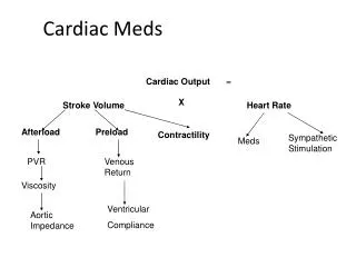
Heart Failure • Major manifestation of cardiac disease • Under 1 year of age due to congenital anomaly • Over 1 year with no congenital anomaly may be due to acquired heart disease
Clinical Manifestations of HF • Systemic Venous Congestion • Weight gain, hepatomegaly, edema, jugular vein distension • Pulmonary Venous Congestion • Tachypnea, dyspnea, cough, wheezes • Compensatory Response • Tachycardia, cardiomegaly, diaphoretic, fatigue, failure to grow
Digoxin Therapy • Digoxin increases the force of the myocardial contraction. • Take an apical pulse with a stethoscope for 1 full minute before every dose of digoxin. If bradycardia is detected. • < 100 beats / min for infant and toddler • < 80 beats in the older child • < 60 beats in the adolescent * Call physician before administering the drug.
Signs of Digoxin Toxicity • Bradycardia • Arrhythmia • Nausea, vomiting, anorexia • Dizziness, headache • Weakness and fatigue
Interventions • Fluid restriction • Diuretics – Lasix (potassium wasting) or Aldactone (potassium sparing) • Bed rest • Oxygen • Small frequent feedings – soft nipple with supplemental NG for adequate calorie intake • Pulse oximeter • Sedatives if needed
Feeding • Small frequent feedings • Soft nipple to easy energy needed to suck • 24 calorie formula for added calories • NG feed if not taking in adequate calories to gain weight
Cardiac Heart Defects • http://www.cincinnatichildrens.org/health/heart-encyclopedia/anomalies/
Patent Ductus Arteriosus • PDA • Incidence 10% • One of the most common benign defects • Ductus normally closes within hours of birth • Connection between the pulmonary artery (low pressure) and aorta (high pressure) • High risk for pulmonary hypertension
Diagnosis and Treatment • Diagnosis by • Chest x-ray – enlarged heart and dilated pulmonary artery • Echo-cardiogram – show the opening between pulmonary artery and aorta
Treatment • Indomethocin given po – constricts the muscle in the wall of the PDA and promotes closure • Cardiac Catheterization – coil is placed in the open duct and acts like a plug • Closed heart surgery – small incision made between ribs on left hand side and PDA is ligated or tied and cut
Atrial Septal Defect • ASD • 10% of defects • Blood in left atrium flows into right atrium • Pulmonary hypertension • Reduced blood volume in systemic circulation • If left untreated may lead to pulmonary hypertension, congestive heart failure or stroke as an adult.
Diagnosis and Treatment • Diagnosis: heart murmur may be heard in the pulmonary valve area because the heart is forcing an unusually large amount of blood through a normal sized valve. • Echocardiogram is the primary method used to diagnose the defect – it can show the hole and its size and any enlargement of the right atrium and ventricle in response to the extra work they are doing.
Treatment • Surgical closure of the atrial septal defect • After closure in childhood the heart size will return to normal over a period of four to six months. • No restrictions to physical activity post closure