CARDIAC MUSCLE
CARDIAC MUSCLE. Dr. Ayisha Qureshi Assistant Professor, MBBS, MPhil. Location of the Heart:. The Pericardium & the Pericardial Sac: .


CARDIAC MUSCLE
E N D
Presentation Transcript
CARDIAC MUSCLE Dr. Ayisha Qureshi Assistant Professor, MBBS, MPhil
The Pericardium & the Pericardial Sac: The heart is enclosed in the double-walled, membranous pericardial sac (perimeans “around”). The sac consists of two layers—a tough, fibrous covering and a secretory lining. The outer fibrous covering of the sac attaches to the connective tissue partition that separates the lungs. This attachment anchors the heart so that it remains properly positioned within the chest. The sac’s secretory lining secretes a thin pericardial fluid, which provides lubrication to prevent friction between the pericardial layers as they glide over each other with every beat of the heart. Pericarditis, an inflammation of the pericardial sac that results in a painful friction rub between the two pericardial layers, occurs occasionally because of viral or bacterial infection.
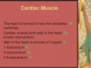
Heart walls are composed of 3 distinct layers: • ENDOCARDIUM (inner): thin layer of endothelium that lines the entire circulatory system (endomeans “within”) • MYOCARDIUM (middle): composed of cardiac muscle that forms the bulk of heart wall (myo means “muscle”) • EPICARDIUM (outer): thin external membrane covering the heart (epi means “on”)
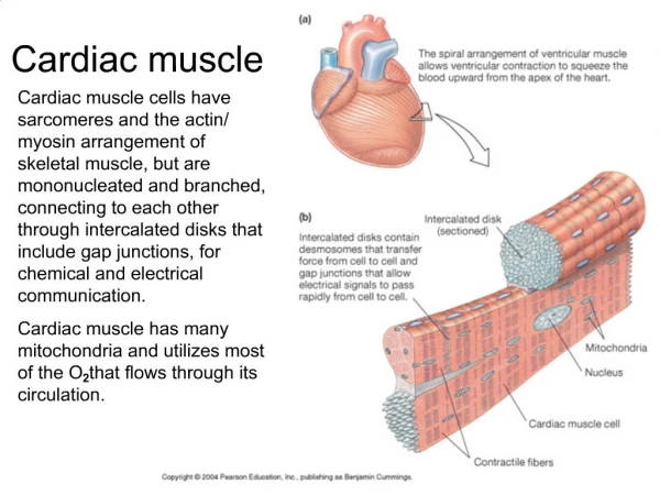
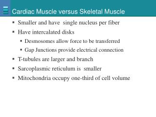
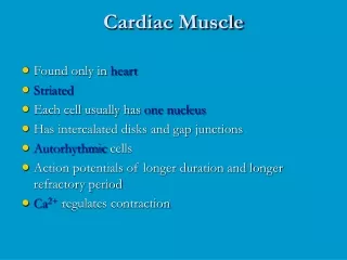
The Cardiac Muscle Branched Centrally located nucleus Muscle cells of the heart-more commonly called myocytes or myofibrils. Outside membrane is called sarcolemma. Cardiac muscles have the same arrangement of actin and myosin, and the same bands, zones and Z discs as skeletal muscles forming sarcomeres. They do have less sarcoplasmic reticulum than skeletal muscles and require Calcium from extra cellular fluid for contraction as T-tubules are not well organized.
Arrangement of the heart muscles The myocardium consists of interlacing bundles of cardiac muscle fibers arranged spirally around the circumference of the heart. What is the advantage of the spiral arrangement? When the cardiac muscle contracts & shortens, a wringing effect is produced, efficiently pushing blood upwards towards the exit of the major arteries of the heart.
Intercalated Discs Although the cardiac muscles interdigitate & branch, there is no anatomical continuity b/w the individual muscle fibers. • Cardiac muscle are branched, have a single nucleus and are interconnected to each other, end to end by specialized structures called as INTERCALATED DISCS.The intercalated discs are further composed of: • Gap Junctions • Desmosomes
HEART AS A DUAL PUMP: Even thought the heart is a single organ, the left and the right side of the heart is anatomically and functionally separate. This is done with the help of the interventricularspetum. It ensures that the blood from the left and right side of the heart does not mix. Although the left and the right sides are separated, the heart contracts in a co-ordinated fashion: the atria contract together and the ventricles contract together….
O2 poor blood returns from the body thru the Superior & Inferior Vena Cava ↓ Enters the Right atrium ↓ Right ventricle ↓ Pulmonary artery ↓ Lungs ↓ Blood is Oxygenated ↓ Pulmonary Veins ↓ Left Atrium ↓ Left Ventricle ↓ Aorta ↓ Circulated to the body