Download
1 / 12
150 likes | 443 Vues
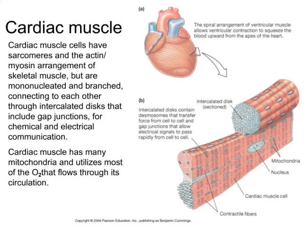
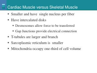
Cardiac Muscle. Prof. K. Sivapalan. Properties of cardiac muscle. Branching cells with central nucleous . Separated by intercalated discs – tight junctions with pores permeable to ions. [electrical continuity] Functional syncytium. Striations – similar to skeletal muscles.

E N D
Cardiac Muscle Prof. K. Sivapalan
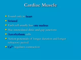
Properties of cardiac muscle. • Branching cells with central nucleous. • Separated by intercalated discs – tight junctions with pores permeable to ions. [electrical continuity] • Functional syncytium. • Striations – similar to skeletal muscles. Cardiac muscle
Electrical properties of cardiac muscle. • Resting membrane potential – 85 – 95 mV. • Depolarized to +20 mV. • Rising phase – 2 m sec. • Plateau – 0.15-0.2 sec in atrium and 0.3 in ventricles. • Refractory period – 0.3 sec. Cardiac muscle
Ionic basis of action potential. • Depolarization – sodium influx. • Plateau – calcium influx and potassium efflux. • Repolarization – potassium efflux. Na+. Ca++ K+. Cardiac muscle
Sarcomere, filaments and fibrils. Z lines – center of actin filaments. • M line – center of myosin filaments. • A band – length of myosin filaments. • Sarcomere is a unit of myofibrils between two Z lines. Cardiac muscle
Myofibrils and T tubular system. • Myofibrils - bundle of actin + myosin [Yellow] • Mitochondria [blue]. • Sarcoplasmic reticulum + T tubules [pink] at Z line. • Intercalated discs at Z line [light blue]. • Central nucleus [purple]. Cardiac muscle
Excitation contraction coupling. • Action potential spreads across intercalated discs. • Spreads along T tubules [Z line] to Terminal cistern. • Calcium released from cistern and influx from ECF. • Actin myosin binding and sliding. • Removal of Calcium results in relaxation. Cardiac muscle
Non-tetanization • The muscle twitch lasts for about 300 ms. • The refractory period extends until more than half of the relaxation period Cardiac muscle
Initial Length and Force • Initial length is proportional to the force of contraction • Starling’s law • Excessive stretch- reduction of force [as in skeletal muscle] Cardiac muscle
Conducting system. • SA node. • Inter nodal pathways & atrial musculature. • AV node. • Bundle of His. • Bundle branches – Purkinje fibers. • Cardiac muscles through intercalated discs. Cardiac muscle
Properties of Conducting System • Pacemaker – junctional tissue. • Pacemaker potential – after each impulse declines to firing level. • Rate of action potential depends on the slope of the prepotential. • It is due to reduction of K+ efflux (↑ by Ach) and then increase in Ca++ influx (↑ by NA). • Ca++ T (transient) channels complete prepotential and L (long lasting) action potentials [no sodium] in nodal tissues. • SA node – 120/min, AV node – 45/min, Purkinje system – 35/min. • First area to reach threshold will be the pace maker. Cardiac muscle
Innervation • No motor end plates- nerves end in varicosities • Sympathetics innervate the nodes and myocardium [noradrenaline] • 10th cranial nerve, vagus, innervates SA and AV nodes of the heart [acetyl choline]. • Stimulation causes chronotropic and ionotropic effects. Cardiac muscle