Download
1 / 96
1.01k likes | 1.17k Vues
Learn about the structure, function, and regulation of cardiac muscle, including action potentials, heart sounds, circulation circuits, cardiac arrhythmias, cardiac cycle, MAP, heart regulation, and homeostasis mechanisms.

E N D
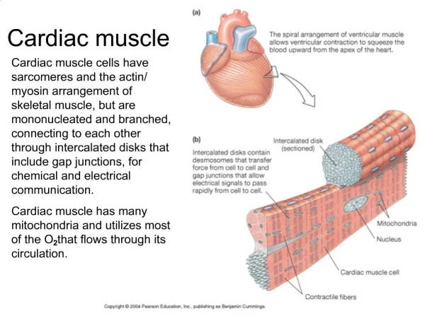
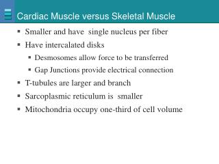
Cardiac Muscle • Found only in heart • Striated • Each cell usually has one nucleus • Has intercalated disks and gap junctions • Autorhythmic cells • Action potentials of longer duration and longer refractory period • Ca2+ regulates contraction
Cardiac Muscle • Elongated, branching cells containing 1-2 centrally located nuclei • Contains actin and myosin myofilaments • Intercalated disks: Specialized cell-cell contacts • Desmosomes hold cells together and gap junctions allow action potentials • Electrically, cardiac muscle behaves as single unit
Refractory Period • Absolute: Cardiac muscle cell completely insensitive to further stimulation • Relative: Cell exhibits reduced sensitivity to additional stimulation • Long refractory period prevents tetanic contractions
AP-contraction relationship: • AP in skeletal muscle is very short-lived • AP is basically over before an increase in muscle tension can be measured. • AP in cardiac muscle is very long-lived • AP has an extra component, which extends the duration. • The contraction is almost over before the action potential has finished.
Functions of the Heart • Generating blood pressure • Routing blood • Heart separates pulmonary and systemic circulations • Ensuring one-way blood flow • Heart valves ensure one-way flow • Regulating blood supply • Changes in contraction rate and force match blood delivery to changing metabolic needs
Orientation of cardiac muscle fibres: • Unlike skeletal muscles, cardiac muscles have to contract in more than one direction. • Cardiac muscle cells are striated, meaning they will only contract along their long axis. • In order to get contraction in two axis, the fibres wrap around.
Heart Wall • Three layers of tissue • Epicardium: This serous membrane of smooth outer surface of heart • Myocardium: Middle layer composed of cardiac muscle cell and responsibility for heart contracting • Endocardium: Smooth inner surface of heart chambers
Heart Sounds • First heart sound or “lubb” • Atrioventricular valves and surrounding fluid vibrations as valves close at beginning of ventricular systole • Second heart sound or “dupp” • Results from closure of aortic and pulmonary semilunar valves at beginning of ventricular diastole, lasts longer • Third heart sound (occasional) • Caused by turbulent blood flow into ventricles and detected near end of first one-third of diastole
Cardiac Arrhythmias • Tachycardia: Heart rate in excess of 100bpm • Bradycardia: Heart rate less than 60 bpm • Sinus arrhythmia: Heart rate varies 5% during respiratory cycle and up to 30% during deep respiration • Premature atrial contractions: Occasional shortened intervals between one contraction and succeeding, frequently occurs in healthy people
Cardiac Cycle • Heart is two pumps that work together, right and left half • Repetitive contraction (systole) and relaxation (diastole) of heart chambers • Blood moves through circulatory system from areas of higher to lower pressure. • Contraction of heart produces the pressure
Mean Arterial Pressure (MAP) • Average blood pressure in aorta • MAP=CO x PR • CO is amount of blood pumped by heart per minute • CO=SV x HR • SV: Stroke volume of blood pumped during each heart beat • HR: Heart rate or number of times heart beats per minute • Cardiac reserve: Difference between CO at rest and maximum CO • PR is total resistance against which blood must be pumped
Regulation of the Heart • Intrinsic regulation: Results from normal functional characteristics, not on neural or hormonal regulation • Starling’s law of the heart • Extrinsic regulation: Involves neural and hormonal control • Parasympathetic stimulation • Supplied by vagus nerve, decreases heart rate, acetylcholine secreted • Sympathetic stimulation • Supplied by cardiac nerves, increases heart rate and force of contraction, epinephrine and norepinephrine released
Heart Homeostasis • Effect of blood pressure • Baroreceptors monitor blood pressure • Effect of pH, carbon dioxide, oxygen • Chemoreceptors monitor • Effect of extracellular ion concentration • Increase or decrease in extracellular K+ decreases heart rate • Effect of body temperature • Heart rate increases when body temperature increases, heart rate decreases when body temperature decreases
Pacemaker regulation: • Once the pacemaker cells reach threshold, the magnitude and duration of the AP is always the same. • In order to change the frequency, the time between APs must vary. • The interval can only be changed in two ways. • The rate of depolarization can be changed • The amount of depolarization required to reach threshold can be changed.
Peripheral Circulatory System • Systemic vessels • Transport blood through most all body parts from left ventricle and back to right atrium • Pulmonary vessels • Transport blood from right ventricle through lungs and back to left atrium • Blood vessels and heart regulated to ensure blood pressure is high enough for blood flow to meet metabolic needs of tissues
Blood Vessel Structure • Arteries • Elastic, muscular, arterioles • Capillaries • Blood flows from arterioles to capillaries • Most of exchange between blood and interstitial spaces occurs across the walls • Blood flows from capillaries to venous system • Veins • Venules, small veins, medium or large veins
Structure of Arteries and Veins • Three layers except for capillaries and venules • Tunica intima (interna) • Endothelium • Tunica media • Vasoconstriction • Vasodilation • Tunica adventitia (externa) • Merges with connective tissue surrounding blood vessels • Note mistake on figure
Structure of Arteries • Elastic or conducting arteries • Largest diameters, pressure high and fluctuates • Muscular or medium arteries • Smooth muscle allows vessels to regulate blood supply by constricting or dilating • Arterioles • Transport blood from small arteries to capillaries
Structure of Veins • Venules and small veins • Tubes of endothelium on delicate basement membrane • Medium and large veins • Valves • Allow blood to flow toward heart but not in opposite direction • Atriovenous anastomoses • Allow blood to flow from arterioles to small veins without passing through capillaries