Download
1 / 44
490 likes | 766 Vues
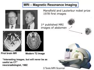
MRI – Magnetic Resonance Imaging. Mansfield and Lauterbur nobel prize 1978 first images. 1 st published MRI images of abdomen. First brain MR. Modern T2 image. “Interesting images, but will never be as useful as CT” neuroradiologist, 1982. 3 Tesla MRI Scanner. MRI.

E N D
MRI – Magnetic Resonance Imaging Mansfield and Lauterbur nobel prize 1978 first images 1st published MRI images of abdomen First brain MR Modern T2 image “Interesting images, but will never be as useful as CT” neuroradiologist, 1982 3 Tesla MRI Scanner
MRI AdvantagesDisadvantages safe expensive great soft tissue contrast long time many contrast options bad for bones mediocre resolution
CT versus MRI MRI +Excellent grey/white matter contrast & spatial resolution +Better for old hemorrhage (and new with Diffusion?) -Long scan time -Pts cannot have metal devices -Claustrophobia, obesity problems +No radiation - expensive CT +Excellent bone imaging +Excellent new acute hemorrhage detection +Skull fracture, calcified lesion +Short scan time, metal devices allowed -Poor contrast and resolution -Radiation
3 Tesla Magnetic Field (60,000 times Earths field) MRI B0 B0 3 1 2
3 Tesla magnet field MRI Not all the protons line up – thermal energy Protons (hydrogen nuclei act like little magnets) B0 Collective Magnetic Moment of Protons Classical picture of Quantum Phenomenon
Model of Head Coil B0
B0 collective spins Model of Head Coil
end start Collective Magnetic Moment of Protons MRI excite Radio Waves B0
Why precession? magnetic moment spin B0 gravity Just like a top on a table
Model of Head Coil excite 3.0 T 123 MHz B0
Model of Head Coil LISTEN signal we “hear” 3.0 T 123 MHz B0
Body Coil - Gradients Radio Waves 123 MHz MRI excite with slice selection B0 Only excite One Slice
excite 3.1 T 127 MHz 3.0 T 123 MHz 2.9 T 119 MHz Like a swing. Got one of the 3 orthogonal spatial dimensions when we excite. z
Model of Head Coil LISTEN signal we “hear” 3.0 T 123 MHz B0
Image should be Image we get of water container
excite 3.1 T 127 MHz 3.0 T 123 MHz 2.9 T 119 MHz Like a swing. Got one of the 3 orthogonal spatial dimensions when we excite. z
Model of Head Coil LISTEN fast 3.1 T 127 MHz regular 3.0 T 123 MHz signal we “hear” slow 2.9 T 119 MHz Got second of the 3 orthogonal spatial dimensions when we listen. x
Image should be Image we get of water container
excite 3.1 T 127 MHz 3.0 T 123 MHz 2.9 T 119 MHz Like a swing. Got one of the 3 orthogonal spatial dimensions when we excite. z
phase encode (after we excite before we listen) fast 3.1 T 127 MHz regular 3.0 T 123 MHz slow 2.9 T 119 MHz Got second of the 3 orthogonal spatial dimensions when we listen. y
Model of Head Coil LISTEN fast 3.1 T 127 MHz regular 3.0 T 123 MHz signal we “hear” slow 2.9 T 119 MHz Got second of the 3 orthogonal spatial dimensions when we listen. x
Repeat 256 times for a 256x256pixel imageDifferent phase each timescan = 4 minutes
Image should be Image we get of water container
SPIN ECHO SEQUENCE 180 Degree RF Pulse Excite Z Y X Listen correctinggradients TE – echo time TR – repeat time
ContrastT1 weighted – (MPRAGE-anatomical)T2 weighted – (fmri)
Spin Relaxation • Spins do not continue to precess forever • Longitudinal magnetization returns to equilibrium due to spin-lattice interactions – T1 decay • Transverse magnetization is reduced due to both spin-lattice energy loss and local, random, spin dephasing – T2 decay • Additional dephasing is introduced by magnetic field inhomogeneities within a voxel – T2' decay. This can be reversible, unlike T2decay
MR Signal start end T1 Recovery Collective Magnetic Moment of Protons B0 T1 decay – “spins back down” signal we “hear” V Time Typical T1 Graph Time
T2 decay – separation (dephasing) of “collective magnetic moment” sometime after RF excitation Immediately after RF excitation collective magnectic moment individual spins a little time later = separation (dephasing) MR Signal T2 Decay Typical T2 Graph Time
MR Signal MR Signal T2 Decay T1 Recovery Proton Density Contrast 1 s 50 ms TE – echo time TR – repeat time
MR Signal T1 Recovery T1 Contrast MR Signal T2 Decay time 1 s time 50 ms TE – echo time TR – repeat time
MR Signal MR Signal T2 Decay T1 Recovery T2* and T2 Contrast 1 s 50 ms TE – echo time TR – repeat time
Proton Density Weighted Image T1 Weighted Image T2 Weighted Image
Tissue T1 (ms) T2 (ms) Grey Matter (GM) 950 100 White Matter (WM) 600 80 Muscle 900 50 Cerebrospinal Fluid (CSF) 4500 2200 Fat 250 60 Blood 1200 100-200 Properties of Body Tissues MRI has high contrast for different tissue types!