Collagen
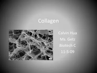
Collagen. Synthesised by VSMC and VEC (perhaps) soluble procollagen excreted cross linked ‘insoluble’ tropocollagen’ assembled into a fibril (details later) fibrils form fibres. Collagen. Most abundant protein in body (25-30% of all protein) >20 distinct forms

Collagen
E N D
Presentation Transcript
Collagen • Synthesised by VSMC and VEC (perhaps) • soluble procollagen excreted • cross linked • ‘insoluble’ tropocollagen’ • assembled into a fibril (details later) • fibrils form fibres
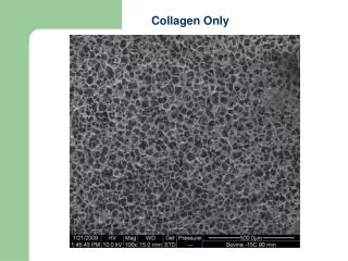
Collagen • Most abundant protein in body (25-30% of all protein) • >20 distinct forms • Types I, III, IV, V, VI, VIII most common in CV system • Types I, III form fibres –major structural elements • synthesized by fibroblasts and SMCs • Type IV forms a porous network (basement membrane) for endothelial cells to attach to (synthesized by ECs) • Types V, VI: provides thin covering over SMCs
Collagen • Fibres are folded at physiologic loads • Half-life in CV system on order of 15-90 days • Degraded by blood borne enzymes • Proteases (matrix metalloproteinases, MMPs) • released by macrophages • De-natured by heat (approx 60°C)
Collagen Fibre Assembly 1 2 3 4 Chains assemble Procollagen peptidase Assembly Assembly 67 nm Precursor chain Procollagen (triple helix with loose ends) Collagen molecule Collagen fibril 10-500 nm Collagen fibre 1-500m Intracellular Stabilised by cross links
Hierarchy of structure Visualisation n.b. 1Å = 0.1nm Size range
Collagen molecule 66nm LH helix 0.87nm RH helix Glycine every third residue
Type I Type II Type III disulphide bonds
Collagen aggregate structure Fibre forming (Types I,II,III,V,K) Membranous (Type IV)
Stagger of collagen molecules in a fibril Total length = 4.4D where D is the length of the striation
Tendon Skin
Ground substance • Mucopolysaccharides (glycosaminoglycans) • Water • Approx 3% of arterial wet weight • Viscous but not elastic
Proteoglycans Proteoglycans • Proteins with covalently bound glycosaminoglycans (linear polymers with disaccharide units) along length • Can hold 50x their dry weight in water • Resistant to compression due to high water content Chondroitin Sulphate Keratin Sulphate Hyaluronate
Fibronectin • Glycoprotein synthesized by fibroblasts, SMCs, ECs • Mediates cellular interactions and migrations (e.g. guides macrophages into wound areas) • Binds fibroblasts to collagen substrates • Crosslinks and stabilizes other components of ECM - • patterns of FN similar to patterns of microfilaments 50m
Laminin • Found primarily in the basal laminae-specialized thin (50 nm) sheets of ECM material separating endothelial cells from ECM • Maintains tissue organization • Acts as “molecular sieve” 10m