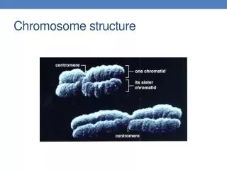
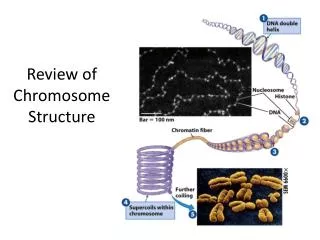
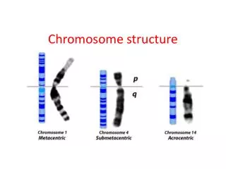
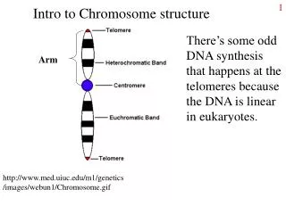
Chromosome Structure Variations
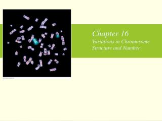
Chromosome Structure Variations. Causes and Problems.

Chromosome Structure Variations
E N D
Presentation Transcript
Causes and Problems • Chromosome structure variations result from chromosome breakage. Broken chromosomes tend to re-join; if there is more than one break, rejoining occurs at random and not necessarily with the correct ends. The result is structural changes in the chromosomes. Chromosome breakage is caused by X-rays, various chemicals, and can also occur spontaneously. • --General problems with structural variants: • 1. breaking a critical gene. This destroys the gene and thus can result in a mutant phenotype. • 2. aneuploidy, usually after meiosis. • We will explore #2 with the individual types of chromosome variation.
Types • --Types: Consider a normal chromosome with genes in alphabetical order: abcdefghi • --deletion: part of the chromosome has been removed: abcghi • --duplication: part of the chromosome is duplicated: abcdefdefghi • --inversion: part of the chromosome has been re-inserted in reverse order: abcfedghi • --ring: the ends of the chromosome are joined together to make a ring • --translocation: parts of two non-homologous chromosomes are joined: if one normal chromosome is abcdefghi and the other chromosome is uvwxyz, then a translocation between them would be abcdefxyz and uvwghi.
Deletions • When homozygous, most deletions are lethal, because most genes are necessary for life and a homozygous deletion would have zero copies of some genes. • When heterozygous, the genes on the normal homologue are hemizygous: there is only 1 copy of those genes, and thus they are expressed even if recessive (like genes on the X in male mammals). • Heterozygous deletions are aneuploid, because the genes in the deleted region are present in only 1 copy instead of the normal two copies. Some genes need to be present in two copies, so heterozygous deletions sometimes give rise to defects in the affected individual, especially if the deletions are large.
Duplications • Genes are duplicated if there is more than one copy present in the haploid genome. • Some duplications are “dispersed”, found in very different locations from each other. • Other duplications are “tandem”, found next to each other. • Tandem duplications play a major role in evolution, because it is easy to generate extra copies of the duplicated genes through the process of unequal crossing over. These extra copies can then mutate to take on altered roles in the cell, or they can become pseudogenes, inactive forms of the gene, by mutation.
Unequal Crossing Over • Unequal crossing over happens during prophase of meiosis 1. Homologous chromosomes pair at this stage, and sometimes pairing occurs between the similar but not identical copies of a tandem duplication. If a crossover occurs within the mispaired copies, one of the resulting gametes will have an extra copy of the duplication and the other will be missing a copy.
Hemoglobin Example • As an example, the beta-globin gene cluster in humans contains 6 genes, called epsilon (an embryonic form), gamma-G, gamma-A (the gammas are fetal forms), pseudo-beta-one (an inactive pseudogene), delta (1% of adult beta-type globin), and beta (99% of adult beta-type globin. Gamma-G and gamma-A are very similar, differing by only 1 amino acid. • If mispairing in meiosis occurs, followed by a crossover between delta and beta, the hemoglobin variant Hb-Lepore is formed. This is a gene that starts out delta and ends as beta. Since the gene is controlled by DNA sequences upstream from the gene, Hb-Lepore is expressed as if it were a delta. That is, it is expressed at about 1% of the level that beta is expressed. Since normal beta globin is absent in Hb-Lepore, the person has severe anemia.
Inversions • An inversion is when a segment of a chromosome is removed and then replaced backwards. • The problem with inversions occurs in meiosis, when a chromosome containing an inversion is heterozygous with a normal chromosome. A crossover within the inverted region results in aneuploidy and death of the resulting embryo. One consequence of this is that crossing over is apparently suppressed; this is seen as a compression of map distances, as you will see in the lab in experiment 2. • Inversions can be either paracentric, where the centromere is NOT in the inverted region, or pericentric, where the inversion is in the inverted region.
Paracentric Inversions • When a paracentric inversion crosses over with a normal chromosome, the resulting chromosomes are an acentric, with no centromeres, and a dicentric, with 2 centromeres. • The acentric chromosome isn't attached to the spindle, so it gets lost during cell division, and the dicentric is usually pulled apart (broken) by the spindle pulling the two centromeres in opposite directions. These conditions are lethal.
Pericentric Inversions • When a pericentric inversion crosses over with a normal chromosome, the resulting chromosomes are both duplicated for some genes and deleted for other genes. (They do have 1 centromere apiece though). The gametes resulting from these are aneuploid and do not survive. • Thus, either kind of inversion has lethal results when it crosses over with a normal chromosome. The only offspring that survive are those that didn't have a crossover. Thus when you count the offspring you only see the non-crossovers, so it appears that crossing over has been suppressed.
Translocations • In a translocation, two different, non-homologous chromosomes are broken and rejoined to each other. All the genes are present, so an individual with a translocation can be completely normal. However, an individual who is heterozygous for a translocation and a set of normal chromosomes can have fertility problems • The problem occurs during meiosis 1, as the result of confusion about how the chromosomes should segregate to opposite poles. • During prophase and metaphase of M1, the homologous chromosomes pair up. Because translocations have pieces of two different chromosomes attached together, they pair up in a cross-shaped configuration, so all the pieces have a partner. This structure is three-dimensional, not flat, and there is ambiguity about which centromeres are attached to which pole of the spindle. • When anaphase occurs, two main possibilities exist: alternate segregation, where centromeres on opposite sides of the cross go to the same pole, and adjacent segregation, where centromeres on the same side of the cross go to the same pole.
Alternate Segregation • In alternate segregation, the centromeres on opposite sides of the cross go to the same pole in anaphase • Alternate segregation results in euploid gametes: half the gametes get both of the normal chromosomes, and the other half of the gametes get both of the translocation chromosomes.
Adjacent Segregation • In adjacent segregation, the centromeres on the same side of the cross go to the same pole. • Adjacent segregation results in aneuploid gametes (which die): each gamete gets one normal chromosome and one translocation chromosome, meaning that some genes are duplicated and others are deleted in each gamete. • Alternate segregation and adjacent segregation occur with about equal frequency, so in a translocation heterozygote about half the gametes are euploid and viable, and the other half are aneuploid and result in a dead embryo.
Translocational Down Syndrome • Most cases of Down syndrome, trisomy-21, are spontaneous. They are caused by non-disjunction which gives an egg or sperm with two copies of chromosome 21. • However, about 5% of Down’s cases are caused by a translocation between chromosome 21 and chromosome 14. These translocational Down’s cases are heritable: several children in the same family can have the disease. • Both chromosome 14 and chromosome 21 are acrocentric, and the short arms contain no essential genes. • Sometimes a translocation occurs that joins the long arms together on one centromere and the short arms on another centromere. In this case the short arm chromosome is usually lost. The individual thus has a normal chromosome 14, a normal chromosome 21, and a translocation chromosome, called t(14;21). • During meiosis, one possible gamete that occurs has both the normal 21 and the t(14;21) in it. When fertilized, the resulting zygote has 2 copies of the important parts of chromosome 14, but 3 copies of chromosome 21: 2 normal copies plus the long arm on the translocation. This zygote develops into a person with Down syndrome.