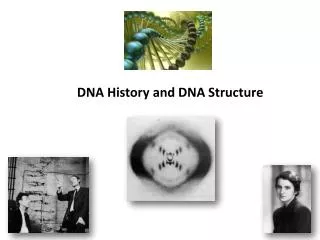
Chromosome Structure and DNA Sequence Organization
540 likes | 1.12k Vues
The wrong file for Lecture 8 was posted on the website. I’ve sent the correct file and it should be posted by the time class is out. Chromosome Structure and DNA Sequence Organization. Bacteria and Viruses.

Chromosome Structure and DNA Sequence Organization
E N D
Presentation Transcript
The wrong file for Lecture 8 was posted on the website. I’ve sent the correct file and it should be posted by the time class is out.
Bacteria and Viruses Bacteria and viruses have only one chromosome, a single piece of DNA that may be linear or looped into a circle. In viruses, the DNA is packed into the phage head. When packaged, the DNA is functionally inert. Once the virus infects a cell, the DNA is released and becomes available for transcription and replication.
Bacterial Chromosome In bacteria, the chromosome is associated with DNA-binding proteins which appear to serve some of the same functions as histones in eukaryotes. Although the DNA is somewhat compacted, it is readily available for transcription and replication.
DNA Packaging in Bacteria Under normal circumstances, DNA in bacteria is supercoiled. To supercoil DNA, the circle is cleaved open, the helix is underwound, then the circle is closed again. When the circle closes, the supercoil forms to restore the proper turn winding of the helix. The resulting structure is very stable, the DNA is available and it takes up much less room.
Supercoiling The enzymes responsible are called topoisomerases. Topoisomerase II: Does supercoils Topoisomerase I: Undoes supercoils
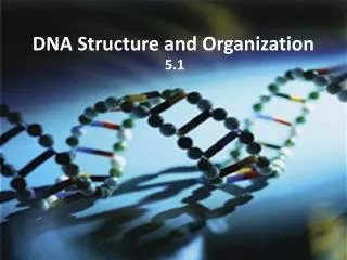
Eukaryotic DNA Organization Eukaryotes have a lot of DNA…. If you hooked all the DNA in all the chromosomes from a single cell, the resulting strand will be two meters in length! That’s a lot of information! To be useful, it must be organized.
Specialized Chromosome Structures Two DNA structures that provided early information about chromosome organization are the Polytene chromosome and the Lampbrush chromosome
Polytene Chromosome Specialized chromosome found in fly larvae. It is actually a pair of homologues, which is unusual (chromatin is the norm)
Polytene Chromosome They are composed of several strands of DNA side by side produced when the DNA undergoes many rounds of replication without separating. The strands present in bands undergo local uncoiling, called a puff. Puffs are areas of high transcriptional activity.
Lampbrush Chromosome Chromosome characteristic of vertebrate oocytes. They are found during the diplotene stage of Prophase I in meiosis. They are synapsed homologue pairs that do not condense like regular chromosomes.
Lampbrush Chromosome They are composed of a center strand (two strands of DNA) with lateral loops (one strand of DNA). The lateral loops are transcriptionally active.
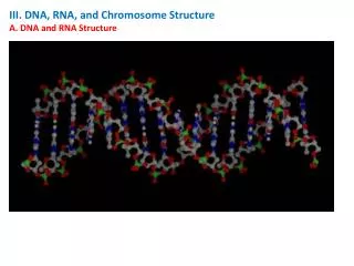
Chromatin Chromatin is the normal form for DNA during Interphase. Chromatin is DNA wrapped around proteins at regular intervals. The most important proteins are called histones.
Histones are responsible for the “beads on a string” appearance of chromatin
Histones are modified to influence gene expression When they are acetylated (an acetyl group attached), the DNA is transcriptionally active. When they are de-acetylated, the DNA is less active. The enzymes responsible are histone acetylases (HAT) and deacetylases (HDAC)
Chromatin Remodeling The degree of acetylation affects chromatin structure, with less tightly wound DNA (more acetylated) being more active. The winding and unwinding of chromatin is referred to as chromatin remodeling.
Histones The stretch of DNA wrapped around a histone is referred to as a nucleosome. A nucleosome contains 150-200 bp DNA
Euchromatin and Heterochromatin During the early days of microscopy, scientists noted that some regions of the chromatin stained darkly, while others stained lightly. Euchromatin is less dense, stains light and is more transcriptionally active. Heterochromatin is much more dense, stains darkly and is much less transcriptionally active.
Heterochromatin Heterochromatic areas are transcriptionally inactive because they either 1. Do not contain genes 2. Contain genes that are repressed Structural components of a chromosome are composed of heterochromatin 1. Centromere regions 2. Telomeres (ends)
Heterochromatin Much of mammalian DNA is heterochromatin. In fact whole chromosomes can be largely heterochromatin (e.g. Y chromosome, the inactivated X chromosome) If a region of euchromatin (active) is translocated to a region of a heterochromatic chromosome, the genes may be inactivated (positional effect).
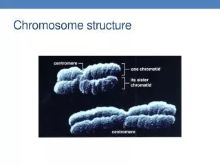
During mitosis and meiosis, chromatin condenses into chromosomes.
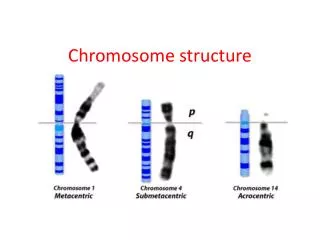
Identifying Chromosome Regions Different staining techniques have been developed to stain particular regions of the chromosomes. The first region to be stained was the center, or centromeric region, creating what are called C-bands.
G-bands The next stain developed produced a banding pattern that allowed researchers to differentiate chromosome pairs when the lengths were similar (like human chromosomes 4 and 5, and 21 and 22
4 5 21 22
Nomenclature When chromosomes are arranged this way, the top arm is called the p arm, and the bottom arm is called the q arm. With the band pattern, genes or traits can be assigned a location on a chromosome Human X chromosome
Sequence Organization within Chromosomes Repetitive DNA Sequences
Repetitive Sequences By far, function of the vast majority of the mammalian genome is unknown. Unique genes (information relating to expression of a protein) only account for about 5% of the genome. Approximately 25% of the mammalian genome is composed of various repetitive DNA sequences.
Repetitive Sequences Various levels of repetition occur throughout the genome, and in numerous places there is considerable variability between individuals in the number of repetitions at a given location.
Repetitive Sequences Highly repetitive sequences are repeated a great number of times and are usually fairly short sequences (10-25 NT). Moderately repetitive sequences are usually tandem repeats or interspersed sequences that are longer than highly repetitive sequences.
The C0t Curve The C0t curve is a measure of the rate at which DNA anneals after it has been denatured to break the hydrogen bonds between individual strands of DNA. The shape of a C0t curve is indicative of how much of DNA is unique vs repetitive. The more repetitive, the faster DNA will reanneal after being denatured by heat. The more unique, the longer it will take for complementary sequences to locate each other and reanneal.
The C0t Curve The C0t curve is the percentage of reassociation of DNA fragments plotted against a log scale of the product of C0 (the initial concentration of DNA strands) and t, or time.
Satellite DNA Satellite DNA is the region on either side of the centromere of each chromosome. When the DNA of chromosomes is denatured the region around the centromere reanneals (bases pair up) much more quickly than the areas farther away from the centromere.
Satellite DNA Satellite DNA is characterized by short sequences repeated many, many times. The repetitive, short sequencces explain why these regions reanneal so quickly: it’s easy to find the match!
Telomeres Telomeres are the ends of the chromosomes, and they function to stabilize the chromosome structure. Telomeres are characterized by many repeats of the sequence 5’GGATT3’. A special enzyme called telomerase synthesizes the very ends of these sequences, to keep telomeres from shrinking during DNA replication.
Telomeres The activity of telomerase varies between cell types. In germ cells (spermatocytes in particular) and cells that undergo continuous cell division, telomerase is very active. In other somatic cells, telomerase activity is limited, so as an organism ages, the chromosomes become progressively shorter, an internal clock of aging.
Moderately Repetitive Sequences There are a number of different moderately repetitive sequences. Minisatellites are variable number tandem repeats (VNTPs). The sequences are variable in length (10-100 bp), but within a repeat sequence, the individual sequences will be the same. VNTPs create regions of 1000-5000 bp in length
Moderately Repetitive Sequences Microsatellites are dinucleotide and trinucleotide repeats that can be repeated up to 50 times. The most common dinucleotide is CA.
Transposable Elements Transposable elements are mobile stretches of DNA that can be moved around from one part of the genome to another. The presence of a transposable element may alter gene expression.
SINES and LINES SINES, or short interspersed elements are only about 200-500 base pairs long but may be present as many as 500,000 times in the human genome. LINES, or long interspersed elements are about 6,000 base pairs long and are present up to 100,000 times in a single genome.