Most Common Contrast Studies in Radiology Basic Principles for interpretation
Most Common Contrast Studies in Radiology Basic Principles for interpretation Robert Cruz, DVM, MS Basic Radiographic Opacities Radiographic image: produced by the aftermath obtained when x ray goes through the body part: penetration and absorption, hence== What you got??

Most Common Contrast Studies in Radiology Basic Principles for interpretation
E N D
Presentation Transcript
Most Common Contrast Studies in RadiologyBasic Principles for interpretation Robert Cruz, DVM, MS
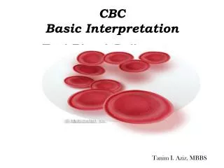
Basic Radiographic Opacities • Radiographic image: produced by the aftermath obtained when x ray goes through the body part: penetration and absorption, hence== What you got?? • Basic radiographic opacities Air Fat Water/ST Bone Metal/+Contrast BLACKGRAY- GRAY - GRAY WHITE
Contrast Radiography • Group of radiographic procedures performed by administration of a contrast medium • Contrast medium: positive (absorb X rays =radiopaque) or negative (do not absorb X – rays, then radiolucent) • What for?? • Visualization of individual organs • Enhance lesions in a particular organ • Some physiologic information • Always performed after a survey radiograph
Contrast Media • Positive • Barium (inert, no hypertonic, no metabolized or absorbed) • Liquid • Paste • Iodine: Tri – iodinated derivatives of benzoic acid • Ionic • Non – ionic • Negative • Air room • Carbon dioxide • Nitrous oxide
Contrast studies for the GI tract • Esophagogram, esophagram, esophogram • Indications: • Suspected esophageal disease • Confirm questionable findings on survey rads • Esophageal motility • Procedure • Survey rads • Sedation not recommended (Acepromazine) • Interpretation • No distention, you can see the bolus • Longitudinal folds and herringbone pattern (cats)
Gastrography • Indications • Suspected gastric wall lesion • Gastric location • Pyloric obstruction (check on fluoroscopy) • Procedure • Double contrast study (barium + air) • Good for gastric mucosa • Single contrast (either barium or air – pneumogastrogram -) • Barium: good for gastric motility • Air: hunt for foreign body • Interpretation • Position • Rugal folds • Motility (gastric emptying) (follow with fluoroscopy/contractions)
Upper GI Study (Series) • Indications • Size, shape, mucosa of SI • Suspect of SI disease • Diagnosis of obstruction • Contraindication • Barium if suspected rupture of hollow viscous, use iodine (ionic) instead • Procedure • Preparation • Interpretation • Lumen diameter • Mucosa
Contrast Studies for the Urinary Tract • Excretory Urography (EU) – Intravenous Pyelogram (IVP) – Intravenous Urography (IVU) • Indications • Evaluate kidney morphology – position. Ureters. • Suspected renal disease • Increased radiopacity in the retroperitoneal space • Gross idea of kidney function • Procedure • Preparation required (24 h fasting; enema) • IV access/dose • Radiographic views
Excretory Urography (EU) – Intravenous Pyelogram (cont’d) • Interpretation • Vascular phase • 5 – 10 sec • Nephrogram phase • 1 – 2 min • Normal pattern: Initial=Good, then=gradual decrease • Abnormal patterns: • Poor – persistent • Poor – continued decrease • Poor – continued increase • Good – continued increase • Pyelogram phase
Cystography • Indications • Suspected bladder disease • Caudal abdominal mass • Bladder rupture • Persistent dysuria /hematuria /stranguria • Congenital anomalies • Procedure • 24 hour fast – enema • Empty bladder • Four views • Positive contrast • Double contrast • Negative contrast (pneumocystogram) • “Paddle” or compression technique
Cystography • Interpretation
Urethrography • Indications • Investigate stranguria / hematuria / dysuria • Urethral rupture • Malformations • Procedure • Positive contrast medium (iodine) • Vaginourethrography • Interpretation
Mielografia • Injeccion de medio de contraste en el espacio subaracnoideo • Medio de contraste: iodado/no ionico: Iopamidol o Iohexol (Omnipaque) • Sito de injeccion: L5 – L6 (mas recomendado), no exito = tratar L6 – L7 => no exito tratar L4 – L5