Download
1 / 73
1.69k likes | 5.14k Vues
Radiology of Fracture Principles. Suzanne O’Hagan 18 May 2012. Radiographic Principles. When analysing and ordering x-rays you should remember the rule of two’s: Two views . At 90 degrees, usually anterior-posterior and lateral. Two joints . The joints above and below.

E N D
Radiology of Fracture Principles Suzanne O’Hagan 18 May 2012
Radiographic Principles • When analysing and ordering x-rays you should remember the rule of two’s: • Two views. At 90 degrees, usually anterior-posterior and lateral. • Two joints. The joints above and below. • Two occasions. Some fractures are not easily visible immediately after trauma. • Two limbs. If required for comparison. • NB: In certain injuries, ‘special’ views are required. These include Scaphoid views, Skyline views for the patello-femoral compartment of the knee and Mortise view at the ankle.
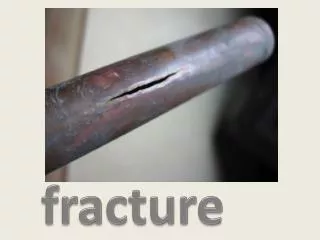
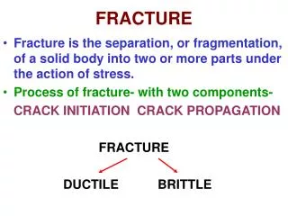
Recognizing an acute fracture Disruption in the continuity of all or part of the cortex of a bone Complete:cortex broken through and through, traversing the width of the bone Incomplete: part of cortex fractured. Tend to occur in bones that are “softer” such as in children or in adults with bone-softening diseases such as osteomalacia or Paget’s disease Examples of incomplete fracture in children are: Greenstick fracture, involves only one part of the cortex - Buckle fracture, compression of cortex
Fracture Lines • More lucent than other lines normally found in bones such as nutrient canals • Abrupt discontinuity of the cortex • Straighter in their course yet more acute in their angulation than naturally occurring lines such as epiphyseal plates • The edges tend to be jagged and rough
Pitfalls • Sesamoids • Bones that form in a tendon as it passes over a joint. The patella is the largest. • Accessory ossicles • These are accessory epiphyseal or apophyseal ossification centres that do not fuse with the parent bone Unlike fractures these small bones are corticated and their edges are usually smooth Sesamoids and accessory ossicles are usually bilateral and at anatomically predictable sites • Old, unhealed fracture fragments • Can be confused with new fractures
Sesamoid bones at joints Knee – the patella (within the quadriceps tendon) Hand– two sesamoid bones commonly found in the distal portions of the first metacarpal bone (within the tendons of adductor pollicis and flexor pollicisbrevis); also distal portion of second metacarpal bone Wrist– the pisiform within the flexor carpiulnaris tendon Foot – first metatarsal bone has two sesamoids at its connection to the big toe, within the tendon of flexor hallucisbrevis (sometimes only a single sesamoid)
Accessory Ossicles • The process of ossification progresses from a primary ossification centre, until the bone is completely ossified. Irregularly shaped bones such as the tarsal bones may develop a secondary centre and in some individuals complete ossification does not occur. The secondary centre remains separate from the rest of the bone, forming an accessory ossicle. Os trigonum – the separated posterolateral tubercle of the talus. Os tibialeexternum (accessory navicular) – located posteromedial aspect of navicular where posterior tibialis tendon inserts
Accessory ossicles • Os Peroneum In peroneusbrevis tendon • Os fabella Posterior to the lateral condyle of the femur. It exists in the location of the lateral head of gastrocnemius tendon. Many more…
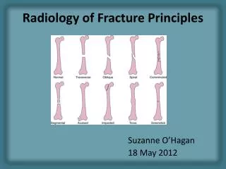
Describing fractures • 4 major parameters • Number of fragments • Direction of fracture line • Relationship of fragments to each other • Communication of the fracture with the outside atmosphere
Number of fracture fragments • 2 fragments = simple fracture • >2 = comminuted fracture • Segmental fracture A portion of the shaft exists as an isolated fragment • Butterfly fragment Central fragment has a triangular shape
Direction of fracture lines • Transverse: Fracture line perpendicular to long axis of bone (perpendicular force) • Oblique: Fracture line diagonal relative to long axis (force usually applied along same direction as long axis) • Spiral: Caused by a twisting force, usually unstable and often associated with soft tissue injury
Relationship of Fragments to each other 1. Displacement 2. Angulation 3. Shortening 4. Rotation • By convention, describe the relationship of the distal fragment relative to theproximal fragment
Displacement • Amount by which the distal fragment is offset, front to back and side to side, from the proximal fragment • Described in terms of percent or fractions (e.g. 50% the width of the shaft or ½ the width of the shaft of the proximal fragment)
Angulation • Angle between the distal and proximal fragments • Described in degrees and by position • State direction of distal bone • Superior, inferior, anterior, posterior, medial, lateral, volar, dorsal • State degree of angulation relative to proximal bone • Medial (varus), Lateral (valgus)
Colles fracture Transverse fracture of distal radius 2.5cm proximal to radiocarpal joint Dorsal displacement and volar angulation
Shortening • How much, if any, overlap there is of the ends of the fracture fragments • How much shorter the fractured bone is than it would be had it not been fractured • Shortening is described in centimetres
Distraction and Impaction • Impaction • – Shortening with no loss • of bone alignment • Distraction • Increase in overall bone length
Rotation • Unusual • Almost always involving the long bones • Describes the orientation of the joint at one end relative to the orientation of the joint at the other end of the fractured bone • Eg proximal tibia oriented in frontal projection while distal tibia and ankle oriented laterally • Both the joint above and below the fracture need to be included to appreciate rotation
Relationship of Fracture to Atmosphere • Closed • More common • No communication • Open/compound • Communication Best diagnosed clinically
Avulsion fractures • Common mechanism of fracture production • Avulsed fragment is pulled from its parent bone by contraction of a tendon or ligament • More common in young athletes • Derive many of their names from athletic activity e.g. dancer’s fracture, skier’s fracture, sprinter’s fracture • Occur in anatomically predictable locations where tendons are known to insert • May heal with exuberant callous formation • Some may resemble a neoplastic or infectious process • Some may have an aggressive appearance that may include areas of mixed lysis and sclerosis • The appearance depends on whether acute, subacute or chronic
Avulsion Fractures: common in the pelvis Avulsion fracture lesser trochanter (iliopsoas) In the pelvis, the newly formed secondary centers of ossification, the apophyses, are most likely to avulse· Apophyses tend to form at the time of puberty = time of pelvic avulsions Avulsion fracture ischialtuberosity (hamstrings)
Don’t confuse with Jone’s Fracture Jones fracture involves a fracture at the base of fifth metatarsal at metaphyseal-diaphyseal junction A Jones fracture is located within 1.5 cm distal to tuberosity of 5th metatarsal Avulsion fracture more common and affects the 5th metatarsal styloid process proximally.
Osgood Schlatter • Caused by stress on the patellar tendon • Patellar tendon attaches the quadriceps muscle to the tibial tuberosity • Adolescent growth spurt, repeated stress from quadriceps contraction is transmitted through the tibial tuberosity • Causes multiple subacute avulsion fractures with inflammation along the tendon leading to excess bone grwoth in the tuberosity
SALTER-HARRIS FRACTURESEpiphyseal plate fractures in children • In growing bone, the hypertrophic zone in the growth plate (epiphyseal plate or physis) is most vulnerable to shearing injuries • Account for as many as 30% of childhood fractures • SH classification helps determine treatment and predict complications • Represent a spectrum of accidental injuries in children
SALTER HARRIS CLASSIFICATION Epiphyseal plate only Epiphyseal plate + metaphysis Epiphyseal plate + epiphysis Compression fracture epiphyseal plate Epiphyseal plate + epiphysis + metaphysis
Prognosis • Types I and II heal well • Type III fractures can develop arthritic changes or asymmetric growth plate fusion • Types IV and V are more likely to develop early fusion of the growth plate with angular deformities and shortening of that bone
Type I: Fractures of the epiphyseal plate alone • Difficult to detect without comparison views • SCFE is a manifestation of a SH I injury • Tall, heavy teenage boys • Bilateral in 25% • Can result in avascular necrosis due to interrupted blood supply in 15%
Salter Harris I “widening of the growth plate” Slipped Capital Femoral Epiphysis
Type II: Fracture of the epiphyseal plate and fracture of metaphysis • Most common (75%) • Seen especially in the distal radius
Salter Harris II “above the growth plate” Distal radius Assoc ulnar fracture
Type III: Fracture of the epiphyseal plate and epiphsysis • Longitudinal fracture through epiphysis itself; fracture invariably enters the joint space and fractures the articular cartilage • Risk of osteoarthritis later in life • Can result in premature and asymmetric fusion of the growth plate with subsequent deformity
Salter Harris III “below the growth plate”
Type IV: Fracture of epiphysis and metaphysis through the epiphyseal plate • Poorer prognosis • premature and possibly asymmetric closure of growth plate • May lead to differences in limb length, angular deformities and secondary OA
Salter Harris IV “through the growth plate”
Salter Harris V • Rare • Associated with vascular injury • Almost always result in growth impairment through early focal fusion of the growth plate • Most common in the distal femur, proximal tibia and distal tibia • Difficult to diagnose on conventional radiographs until later when they complicate
Salter Harris V Right Left
Non-accidental injury patterns • Metaphyseal corner fractures • Rib fractures • Especially multiple and posterior • Head injuries • Skull fractures tend to be bilateral, comminuted and cross suture lines (associated subdurals, SAH, cerebral contusion)
CML: Classic Metaphyseal LesionVirtually pathognomonic of abuse • series of microfractures across the metaphysis • the fracture line is oriented essentially parallel to the physis, although it may not travel the entire width of the bone • precipitating force is a shearing injury across the bone end, the result of horizontal motion across the metaphysis, therefore not a feature of falls or blunt trauma • force is generated by manual to-and-fro manipulation of the extremities (eg, holding and shaking an infant by the feet or hands or shaking the infant while he is held around the chest) • CML is seen almost exclusively in children less than 2 years of age
Stress Fracture • Bone subjected to repeated stretching and compressive forces • Numerous microfractures • Conventional radiographs may initially appear normal in up to 85% • Fracture may not be diagnosable until after periosteal new bone formation or healing occurs • Bone scans or MRI will usually be positive earlier • Common locations include the shafts of long bones, the calcaneus and the 2nd and 3rd metatarsals
5 MOST COMMON EPONYMS • Colle’s • Smith’s • Jones • Boxer’s • March 3 in the hand, 2 in the foot