Paediatric Oncology
300 likes | 1.11k Vues
Paediatric Oncology. Maria Seago, mls57@cam.ac.uk. Aims and Objectives. Know the main types of cancer that affect children Understand how they are diagnosed and treated Recognise and interpret their histopathology. Paediatric Cancers.

Paediatric Oncology
E N D
Presentation Transcript
Paediatric Oncology Maria Seago, mls57@cam.ac.uk
Aims and Objectives • Know the main types of cancer that affect children • Understand how they are diagnosed and treated • Recognise and interpret their histopathology
Paediatric Cancers Very rare – approx. 1800 new cases per year in the UK, according to Cancer Research UK An average GP will see two cases in their career
Paediatric Cancers • ‘Childhood tumours’ • Small round blue cell tumours • Collections of small round cells with little cytoplasm • Often undifferentiated • E.g. Ewing sarcoma, rhabdomyosarcoma, neuroblastoma, Wilms’ tumour
Rhabdomyosarcoma • Tumour of primitive skeletal muscle • Third most common solid tumour in children; 4.5/1 million • Found throughout the body, so presentation can be variable
Rhabdomyosarcoma • Rhabdomyoblasts: eosinophilic primitive myogenic cells (yellow)
Rhabdomyosarcoma • Embryonal • More primitive tumour • More common in young children • Typical presentations include: • GU tract: ‘bunch of grapes’ in bladder or vagina • Pharynx: airway obstruction • Parameningeal/orbital: proptosis • Often localised or with lung mets • Better prognosis • Histology: scattered rhabdomyoblasts and tumour cells set randomly within stroma
Rhabdomyosarcoma • Alveolar • More differentiated tumour • More common in older children • Typical presentations include: • Limb and extremity tumours • Aggressive – may disseminate widely into bone marrow, lungs and brain • Poorer prognosis • Most cases have either a t(1:13) or a t(2:13) translocation involving Pax-7 or Pax-3 and Forkhead (FOXO1A) genes • Histology: collections of cells grow in clusters arranged around a central space, and separated by fibrous septa
Rhabdomyosarcoma • Staging with: • MRI and/or CT of primary site • CT chest to exclude lung mets • Bone scan to look for bony lesions • Bone marrow trephines to look for dissemination • Treatment: • Limb tumours may be successfully cured by amputation • For most rhabdomyosarcomas, surgery followed by radiotherapy +/- chemotherapy is needed, although patients with high-risk disease only have a survival rate of 20-30%
Neuroblastoma • Tumour derived from neural crest cells • Arise within the sympathetic chain, Organ of Zuckerkandl (a chromaffin body located at the bifurcation of the aorta or at the origin of the IMA) and adrenal medulla • Age typically <4 years; ~100 per year in UK
Neuroblastoma • Most typically present as a supra-renal mass. Other presentations may include: • Swollen abdomen • Pain • Failure to thrive • Jaundice/anaemia (from disease spread) • Hypertension • Skin lesions ‘blueberry muffin baby’ from haematopoiesis in the skin and soft tissues • Calcification/necrotic mass on CT • Elevated urinary catecholamines (VMA and HVA) in 90% of neuroblastomas. De-differentiated tumours secrete more HVA than VMA.
Neuroblastoma • Staging: • CT scan of primary site • CT chest to look for mets • Head MRI • MIBG scan to look for sites of uptake (radioactive iodine-123 meta-iodobenzylguanidine) • Bone marrow trephines to look for disseminated disease • Prognosis is very variable, and depends on age (congenital neuroblastoma can spontaneously regress), stage, histology (better differentiation = better prognosis) and genetics: • MYCN amplification destabilises p53 and carries a poor prognosis • 80% have chromosome 17q gain, which leads to a poorer outcome (although gain of the whole of C17 has a better outcome) • 30% have chromosome 1p loss, which increases chance of recurrence • 20% have chromosome 11q loss which reduce time of progression free survival • The ALK protein kinase oncogene is frequently responsible for familial neuroblastoma and is involved in 8% of sporadic cases
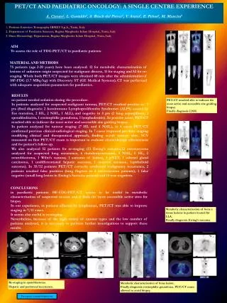
Neuroblastoma • Neuroblasts: small round blue cells. May be arranged in Homer-Wright rosettes with a centre of neuropil. • Neuropil: eosinophilic fibrillary acellular material • Ganglion cells: mature neuroblasts, with abundant pink cytoplasm and a large nucleus with a prominent nucleolus • Schwannian stroma: cellular wavy stroma formed from Schwann cells
Neuroblastoma • Ganglioneuroma • Most mature • >50% Schwannian stroma • Ganglion cells present • No neuroblasts • Neuroblastoma • Most immature • <50% Schwannian stroma • Undifferentiated form • No neuropil • Poorly differentiated form • Some neuropil • <5% ganglion cells • Differentiating form • Neuropil • >5% ganglion cells • Ganglioneuroblastoma • More mature • >50% Schwannian stroma • Neuroblasts present, either as nodules (nodular GNB) or scattered (intermixed GNB) • Ganglion cells present
Neuroblastoma Ganglioneuroblastoma Ganglioneuroma
Wilms’ Tumour • Tumour of primitive renal tissue • Usually <6 years but not neonates: • 84% renal masses in childhood are Wilms’ tumour • But if under 3 months, mesoblastic nephroma (a benign mesenchymal tumour) accounts for 66% of renal masses • Presentation is typically: • Abdominal mass • Hypertension • Intra-tumour haemorrhage, often causing pain and/or haematuria
Wilms’ Tumour Histological staging is from analysis of the resected kidney. Stage 1 = isolated to kidney Stage 2 = infiltration of renal sinus Stage 3 = At margins or in lymph nodes Stage 4 = metastasis • Staging: • CT and (sometimes) MRI of the abdomen • CT chest to look for lung mets • Bone marrow assessment NOT done as Wilms’ tumour does not typically spread to the marrow • 17% of cases are syndromic: • WT-1 deletions including WAGR syndrome • WT-1 truncation and pathogenic missense mutations including Denys-Drash Syndrome • Familial Wilms tumour • Perlman Syndrome • Fanconi Anaemia D1 • Mosaic variegated aneuploidy
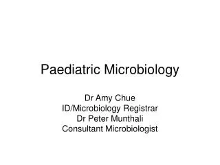
Wilms’ Tumour • Histologically they typically have three components: • Epithelium: tubules, papillary structures, glomerular structures • Blastema: sheets of small round blue cells • Stroma: fibrous/myxoid tissue
Wilms’ Tumour • Anaplastic Wilms’ tumour has a poor prognosis: • Nuclei 3 x size of normal tumour nuclei • Nuclei hyperchromatic • Abnormal mitoses
Wilms’ Tumour • Nephrogenic rests are precursors of Wilms’ tumour: • Foci of abnormally persistent embryonal tissue after 36 weeks gestation • 1% paediatric kidneys; 35% unilateral WT; 100% bilateral WT • May be intralobar or perilobar. • Intralobar rests have an increased rate of progression to Wilms tumor, are more commonly associated with WT1 mutations, Denys-Drash syndrome and WAGR syndrome • Perilobar are associated with genetic / epigenetic dysregulation at 11p15, idiopathic hemihypertrophy and Beckwith-Wiedemann syndrome
Burkitt Lymphoma High grade B cell lymphoma characterized by CD10+ and c-MYC translocation • Endemic • Endemic in Equatorial Africa and Papua New Guinea • Mostly affects children aged 4-7 years • Associated with malaria, EBV+ • Immunodeficiency-associated • HIV-associated • 25-40% EBV+ • Appears early when CD4 counts are still high • Sporadic • Worldwide • Mostly affects children and young adults • Comprises 1-2% of lymphomas (but 30-50% of paediatric lymphomas) • 30% are EBV+ • Also associated with low socioeconomic status
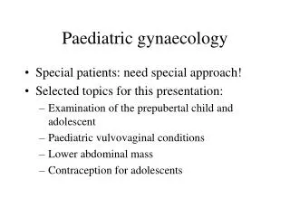
Burkitt Lymphoma Macrophages phagocytizing apoptotic cell debris
Ewing’s Sarcoma • Tumour of primitive neuroectoderm, hence may involve bone and soft tissue throughout the body • Typically presents with: • Bone pain • Pathological fracture • Swelling • Pelvic mass with obstruction and spinal cord encroachment • Symptoms from metastases
Ewing’s Sarcoma • Histology: • Small round blue cells • May be biphasic with dark/light areas • May see rosettes • Membranous staining for CD99 (MIC) • Molecularly: • 95% have a translocation involving the EWS gene on chromosome 22 • Commonest translocation partner is FLI-1 on chromosome 11
Retinoblastoma • Usually affects children under the age of 5 • Around 45 in the UK per year • About 40% have a heritable type, which often affects both eyes. This is caused by the RB1 tumour suppressor gene on chromosome 13. Usually it is passed on from a parent and the allele has 90% penetrance, but it may be a de novo mutation • Somatic amplification of the MYCN oncogene is responsible for some cases of non-hereditary, early-onset, aggressive, unilateral retinoblastoma.
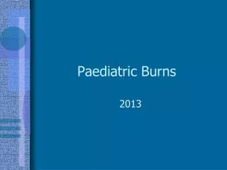
Retinoblastoma Proptosis and exomphalos in developing countries Red reflex showing leukocoria Hirschberg test for strabismus
Retinoblastoma • Differentials: • Cataract • Coat’s disease (rare congenital, nonhereditary eye disorder, causing full or partial blindness, characterized by abnormal development of blood vessels behind the retina) • Retinopathy of prematurity • Vitreous haemorrhage • Other retinal tumours such as astrocytic hamartoma
Retinoblastoma • Treatment: • High cure rate of ~98% • Enucleation if the definitive treatment and very successful • To preserve vision: systemic chemotherapy with focal consolidation, intra-arterial chemotherapy, and for small tumours, focally destructive therapy (cryopexy, laser photocoagulation, hyperthermia and plaque irradiation). • Radiotherapy may really be needed • Children who have a parent or brother or sister who had retinoblastoma should be checked for retinoblastoma. They usually have screening from birth to the age of 3 years. This involves regular eye examinations under a general anaesthetic
Thank you! • https://goo.gl/forms/aqF6hYvqz3GYebXi2 • Check out: https://vle.medschl.cam.ac.uk/mod/scorm/player.php?a=77¤torg=XERTE-ORG-1518514574&scoid=163&sesskey=q7arCtIYRP&display=popup&mode=normal